5DIL

Crystal structure of the effector domain of the NS1 protein from influenza virus B


Experimental Data Snapshot

  • Method: X-RAY DIFFRACTION
  • Resolution: 2.01 Å
  • R-Value Free: 0.242 
  • R-Value Work: 0.194 
  • R-Value Observed: 0.199 

wwPDB Validation   3D Report Full Report


This is version 1.3 of the entry. See complete history


Literature

A Second RNA-Binding Site in the NS1 Protein of Influenza B Virus.

Ma, L.C.Guan, R.Hamilton, K.Aramini, J.M.Mao, L.Wang, S.Krug, R.M.Montelione, G.T.

(2016) Structure 24: 1562-1572

  • DOI: https://doi.org/10.1016/j.str.2016.07.001
  • Primary Citation of Related Structures:  
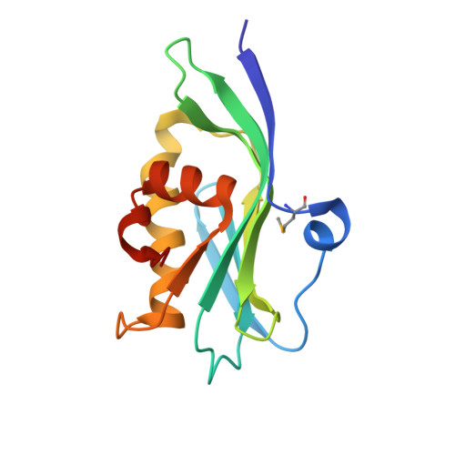
    5DIL

  • PubMed Abstract: 

    Influenza viruses cause a highly contagious respiratory disease in humans. The NS1 proteins of influenza A and B viruses (NS1A and NS1B proteins, respectively) are composed of two domains, a dimeric N-terminal domain and a C-terminal domain, connected by a flexible polypeptide linker. Here we report the 2.0-Å X-ray crystal structure and nuclear magnetic resonance studies of the NS1B C-terminal domain, which reveal a novel and unexpected basic RNA-binding site that is not present in the NS1A protein. We demonstrate that single-site alanine replacements of basic residues in this site lead to reduced RNA-binding activity, and that recombinant influenza B viruses expressing these mutant NS1B proteins are severely attenuated in replication. This novel RNA-binding site of NS1B is required for optimal influenza B virus replication. Most importantly, this study reveals an unexpected RNA-binding function in the C-terminal domain of NS1B, a novel function that distinguishes influenza B viruses from influenza A viruses.


  • Organizational Affiliation

    Department of Molecular Biology and Biochemistry, Center for Advanced Biotechnology and Medicine, Rutgers, The State University of New Jersey, Northeast Structural Genomics Consortium, Piscataway, NJ 08854, USA.


Macromolecules
Find similar proteins by:  (by identity cutoff)  |  3D Structure
Entity ID: 1
MoleculeChains Sequence LengthOrganismDetailsImage
Non-structural protein 1
A, B
141Influenza B virus (B/Singapore/DSO_090134/2004)Mutation(s): 0 
Gene Names: NS1
UniProt
Find proteins for X2C382 (Influenza B virus)
Explore X2C382 
Go to UniProtKB:  X2C382
Entity Groups  
Sequence Clusters30% Identity50% Identity70% Identity90% Identity95% Identity100% Identity
UniProt GroupX2C382
Sequence Annotations
Expand
  • Reference Sequence
Small Molecules
Ligands 1 Unique
IDChains Name / Formula / InChI Key2D Diagram3D Interactions
IOD
Query on IOD

Download Ideal Coordinates CCD File 
C [auth A]
D [auth A]
E [auth A]
F [auth A]
G [auth A]
C [auth A],
D [auth A],
E [auth A],
F [auth A],
G [auth A],
H [auth A],
I [auth A],
J [auth A],
K [auth B],
L [auth B],
M [auth B],
N [auth B],
O [auth B]
IODIDE ION
I
XMBWDFGMSWQBCA-UHFFFAOYSA-M
Modified Residues  1 Unique
IDChains TypeFormula2D DiagramParent
MSE
Query on MSE
A, B
L-PEPTIDE LINKINGC5 H11 N O2 SeMET
Experimental Data & Validation

Experimental Data

  • Method: X-RAY DIFFRACTION
  • Resolution: 2.01 Å
  • R-Value Free: 0.242 
  • R-Value Work: 0.194 
  • R-Value Observed: 0.199 
  • Space Group: P 1
Unit Cell:
Length ( Å )Angle ( ˚ )
a = 34.689α = 68.72
b = 41.666β = 82.83
c = 49.681γ = 78.12
Software Package:
Software NamePurpose
PHENIXrefinement
HKL-2000data reduction
HKL-2000data scaling
PHENIXphasing

Structure Validation

View Full Validation Report


Entry History & Funding Information

Deposition Data


Funding OrganizationLocationGrant Number
National Institutes of Health/National Institute of General Medical Sciences (NIH/NIGMS)United States--

Revision History  (Full details and data files)

  • Version 1.0: 2016-08-10
    Type: Initial release
  • Version 1.1: 2016-10-05
    Changes: Database references
  • Version 1.2: 2017-09-27
    Changes: Author supporting evidence, Database references, Derived calculations
  • Version 1.3: 2019-12-25
    Changes: Author supporting evidence