5C2N

The de novo evolutionary emergence of a symmetrical protein is shaped by folding constraints


Experimental Data Snapshot

  • Method: X-RAY DIFFRACTION
  • Resolution: 1.65 Å
  • R-Value Free: 0.248 
  • R-Value Work: 0.186 
  • R-Value Observed: 0.189 

wwPDB Validation   3D Report Full Report


Ligand Structure Quality Assessment 


This is version 1.3 of the entry. See complete history


Literature

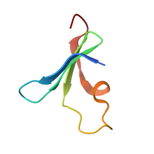
De Novo Evolutionary Emergence of a Symmetrical Protein Is Shaped by Folding Constraints.

Smock, R.G.Yadid, I.Dym, O.Clarke, J.Tawfik, D.S.

(2016) Cell 164: 476-486

  • DOI: https://doi.org/10.1016/j.cell.2015.12.024
  • Primary Citation of Related Structures:  
    5C2M, 5C2N

  • PubMed Abstract: 

    Molecular evolution has focused on the divergence of molecular functions, yet we know little about how structurally distinct protein folds emerge de novo. We characterized the evolutionary trajectories and selection forces underlying emergence of β-propeller proteins, a globular and symmetric fold group with diverse functions. The identification of short propeller-like motifs (<50 amino acids) in natural genomes indicated that they expanded via tandem duplications to form extant propellers. We phylogenetically reconstructed 47-residue ancestral motifs that form five-bladed lectin propellers via oligomeric assembly. We demonstrate a functional trajectory of tandem duplications of these motifs leading to monomeric lectins. Foldability, i.e., higher efficiency of folding, was the main parameter leading to improved functionality along the entire evolutionary trajectory. However, folding constraints changed along the trajectory: initially, conflicts between monomer folding and oligomer assembly dominated, whereas subsequently, upon tandem duplication, tradeoffs between monomer stability and foldability took precedence.


  • Organizational Affiliation

    Department of Biological Chemistry, Weizmann Institute of Science, Rehovot 76100, Israel.


Macromolecules
Find similar proteins by:  (by identity cutoff)  |  3D Structure
Entity ID: 1
MoleculeChains Sequence LengthOrganismDetailsImage
Beta propeller
A, B, C, D, E
A, B, C, D, E, F, G, H, I, J, K, L, M, N, O
48Enterobacteria phage L1Mutation(s): 0 
UniProt
Find proteins for A0A140UHM9 (Enterobacteria phage L1)
Explore A0A140UHM9 
Go to UniProtKB:  A0A140UHM9
Entity Groups  
Sequence Clusters30% Identity50% Identity70% Identity90% Identity95% Identity100% Identity
UniProt GroupA0A140UHM9
Sequence Annotations
Expand
  • Reference Sequence
Small Molecules
Ligands 1 Unique
IDChains Name / Formula / InChI Key2D Diagram3D Interactions
NAG
Query on NAG

Download Ideal Coordinates CCD File 
AA [auth L]
BA [auth M]
CA [auth M]
DA [auth O]
P [auth A]
AA [auth L],
BA [auth M],
CA [auth M],
DA [auth O],
P [auth A],
Q [auth B],
R [auth C],
S [auth D],
T [auth E],
U [auth E],
V [auth F],
W [auth F],
X [auth I],
Y [auth J],
Z [auth L]
2-acetamido-2-deoxy-beta-D-glucopyranose
C8 H15 N O6
OVRNDRQMDRJTHS-FMDGEEDCSA-N
Experimental Data & Validation

Experimental Data

  • Method: X-RAY DIFFRACTION
  • Resolution: 1.65 Å
  • R-Value Free: 0.248 
  • R-Value Work: 0.186 
  • R-Value Observed: 0.189 
  • Space Group: P 31 1 2
Unit Cell:
Length ( Å )Angle ( ˚ )
a = 112.219α = 90
b = 112.219β = 90
c = 107.29γ = 120
Software Package:
Software NamePurpose
REFMACrefinement
HKL-2000data reduction
SCALEPACKdata scaling
PHASERphasing

Structure Validation

View Full Validation Report


Ligand Structure Quality Assessment 


Entry History 

Deposition Data

Revision History  (Full details and data files)

  • Version 1.0: 2016-01-20
    Type: Initial release
  • Version 1.1: 2016-02-03
    Changes: Database references
  • Version 1.2: 2016-02-10
    Changes: Database references
  • Version 1.3: 2020-07-29
    Type: Remediation
    Reason: Carbohydrate remediation
    Changes: Data collection, Derived calculations, Structure summary