5B4N

Structure analysis of function associated loop mutant of substrate recognition domain of Fbs1 ubiquitin ligase


Experimental Data Snapshot

  • Method: X-RAY DIFFRACTION
  • Resolution: 2.30 Å
  • R-Value Free: 0.266 
  • R-Value Work: 0.185 
  • R-Value Observed: 0.189 

wwPDB Validation   3D Report Full Report


This is version 1.2 of the entry. See complete history


Literature

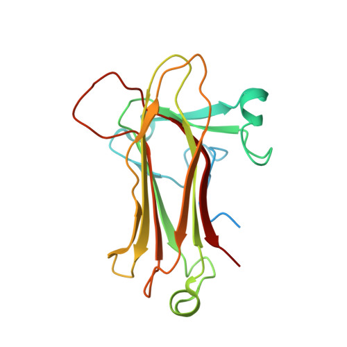
Structural analysis of a function-associated loop mutant of the substrate-recognition domain of Fbs1 ubiquitin ligase

Nishio, K.Yoshida, Y.Tanaka, K.Mizushima, T.

(2016) Acta Crystallogr F Struct Biol Commun 72: 619-626

  • DOI: https://doi.org/10.1107/S2053230X16011018
  • Primary Citation of Related Structures:  
    5B4N

  • PubMed Abstract: 

    The SCF ubiquitin ligase comprises four components: Skp1, Cul1, Rbx1 and a variable-subunit F-box protein. The F-box protein Fbs1, which recognizes the N-linked glycoproteins, is involved in the endoplasmic reticulum-associated degradation pathway. Although FBG3, another F-box protein, shares 51% sequence identity with Fbs1, FBG3 does not bind glycoproteins. To investigate the sequence-structure relationship of the substrate-binding pocket, the crystal structure of a mutant substrate-binding domain of Fbs1 in which the six nonconserved regions (β1, β2-β3, β3-β4, β5-β6, β7-β8 and β9-β10) of Fbs1 were substituted with those of FBG3 was determined. The substrate-binding pocket of this model exhibits structural features that differ from those of Fsb1.


  • Organizational Affiliation

    Picobiology Institute, Graduate School of Life Science, University of Hyogo, 3-2-1 Kouto, Kamigori-cho, Ako-gun, Hyogo 678-1297, Japan.


Macromolecules
Find similar proteins by:  (by identity cutoff)  |  3D Structure
Entity ID: 1
MoleculeChains Sequence LengthOrganismDetailsImage
F-box only protein 2
A, B
203Mus musculusMutation(s): 40 
Gene Names: Fbxo2Fbs1Fbx2
UniProt & NIH Common Fund Data Resources
Find proteins for Q80UW2 (Mus musculus)
Explore Q80UW2 
Go to UniProtKB:  Q80UW2
IMPC:  MGI:2446216
Find proteins for Q9H4M3 (Homo sapiens)
Explore Q9H4M3 
Go to UniProtKB:  Q9H4M3
PHAROS:  Q9H4M3
GTEx:  ENSG00000132879 
Entity Groups  
Sequence Clusters30% Identity50% Identity70% Identity90% Identity95% Identity100% Identity
UniProt GroupsQ80UW2Q9H4M3
Sequence Annotations
Expand
  • Reference Sequence
Experimental Data & Validation

Experimental Data

  • Method: X-RAY DIFFRACTION
  • Resolution: 2.30 Å
  • R-Value Free: 0.266 
  • R-Value Work: 0.185 
  • R-Value Observed: 0.189 
  • Space Group: P 1 21 1
Unit Cell:
Length ( Å )Angle ( ˚ )
a = 44.541α = 90
b = 96.203β = 101.9
c = 44.681γ = 90
Software Package:
Software NamePurpose
REFMACrefinement
HKL-2000data reduction
HKL-2000data scaling
MOLREPphasing

Structure Validation

View Full Validation Report


Entry History & Funding Information

Deposition Data


Funding OrganizationLocationGrant Number
Ministry of Education, Culture, Sports, Science and Technology of JapanJapan24112009

Revision History  (Full details and data files)

  • Version 1.0: 2016-09-14
    Type: Initial release
  • Version 1.1: 2017-08-30
    Changes: Data collection, Derived calculations
  • Version 1.2: 2023-11-08
    Changes: Data collection, Database references, Refinement description