5AFQ

Crystal structure of RPC62 - RPC32 beta


Experimental Data Snapshot

  • Method: X-RAY DIFFRACTION
  • Resolution: 7.00 Å
  • R-Value Free: 0.298 
  • R-Value Work: 0.268 
  • R-Value Observed: 0.271 

wwPDB Validation   3D Report Full Report


This is version 1.2 of the entry. See complete history


Literature

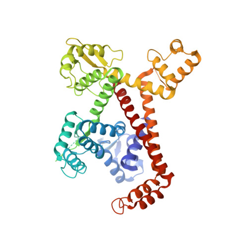
Structural Analysis of Human Rpc32Beta - Rpc62 Complex.

Boissier, F.Dumay-Odelot, H.Teichmann, M.Fribourg, S.

(2015) J Struct Biol 192: 313

  • DOI: https://doi.org/10.1016/j.jsb.2015.09.004
  • Primary Citation of Related Structures:  
    5AFQ

  • PubMed Abstract: 

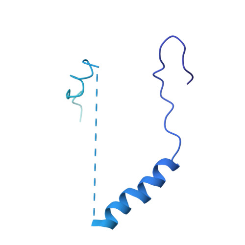
    Transcription initiation by eukaryotic RNA polymerase (Pol) III relies on the subcomplex RPC62/RPC39/RPC32. Two distinct isoforms of RPC32 are encoded in the human genome. RPC32α expression is highly regulated and found only in stem cells and transformed cells, whereas RPC32β is ubiquitously expressed in tissues. Here we identify a core-interacting domain of RPC32 sufficient for the interaction with RPC62. We present the crystal structure of a complex of RPC62 and the RPC32β core domain. RPC32β associates with the extended winged helix 1 and 2 and the coiled coil domain of RPC62 qualifying RPC32 as a molecular bridge in between RPC62 domains. The RPC62-RPC32 complex fit into EM data suggests a bi-functional role for RPC32 through interactions with the largest Pol III subunit and through solvent exposed residues. RPC32 positioning into Pol III suggests that subunit-specific contacts at the surface of the Pol III holoenzyme are critical for its function.


  • Organizational Affiliation

    Université de Bordeaux, Institut Européen de Chimie et Biologie, ARNA Laboratory, F-33607 Pessac, France; Institut National de la Santé Et de la Recherche Médicale, INSERM - U869, ARNA Laboratory, F-33000 Bordeaux, France.


Macromolecules
Find similar proteins by:  (by identity cutoff)  |  3D Structure
Entity ID: 1
MoleculeChains Sequence LengthOrganismDetailsImage
DNA-DIRECTED RNA POLYMERASE III SUBUNIT RPC3
A, B
534Homo sapiensMutation(s): 0 
UniProt & NIH Common Fund Data Resources
Find proteins for Q9BUI4 (Homo sapiens)
Explore Q9BUI4 
Go to UniProtKB:  Q9BUI4
PHAROS:  Q9BUI4
GTEx:  ENSG00000186141 
Entity Groups  
Sequence Clusters30% Identity50% Identity70% Identity90% Identity95% Identity100% Identity
UniProt GroupQ9BUI4
Sequence Annotations
Expand
  • Reference Sequence
Find similar proteins by:  (by identity cutoff)  |  3D Structure
Entity ID: 2
MoleculeChains Sequence LengthOrganismDetailsImage
RPC32 BETA (RPC7L)C [auth D],
D [auth E]
218Homo sapiensMutation(s): 0 
Sequence Annotations
Expand
  • Reference Sequence
Experimental Data & Validation

Experimental Data

  • Method: X-RAY DIFFRACTION
  • Resolution: 7.00 Å
  • R-Value Free: 0.298 
  • R-Value Work: 0.268 
  • R-Value Observed: 0.271 
  • Space Group: I 4 2 2
Unit Cell:
Length ( Å )Angle ( ˚ )
a = 218.27α = 90
b = 218.27β = 90
c = 182.93γ = 90
Software Package:
Software NamePurpose
BUSTERrefinement
MOSFLMdata reduction
SCALAdata scaling
PHASERphasing

Structure Validation

View Full Validation Report


Entry History 

Deposition Data

  • Released Date: 2015-10-07 
  • Deposition Author(s): Fribourg, S.

Revision History  (Full details and data files)

  • Version 1.0: 2015-10-07
    Type: Initial release
  • Version 1.1: 2015-12-02
    Changes: Database references
  • Version 1.2: 2024-01-10
    Changes: Data collection, Database references, Other, Refinement description