5OCY

Crystal structure of ACPA E4 in complex with CII-C-48-CIT


Experimental Data Snapshot

  • Method: X-RAY DIFFRACTION
  • Resolution: 2.60 Å
  • R-Value Free: 0.245 
  • R-Value Work: 0.187 
  • R-Value Observed: 0.190 

wwPDB Validation   3D Report Full Report


This is version 3.1 of the entry. See complete history


Literature

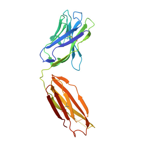
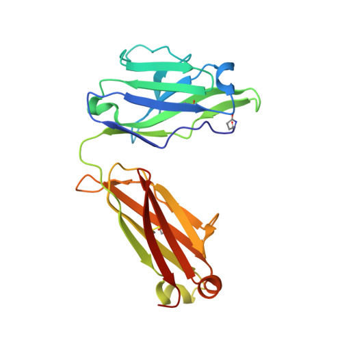
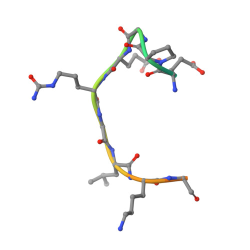
Structural Basis of Cross-Reactivity of Anti-Citrullinated Protein Antibodies.

Ge, C.Xu, B.Liang, B.Lonnblom, E.Lundstrom, S.L.Zubarev, R.A.Ayoglu, B.Nilsson, P.Skogh, T.Kastbom, A.Malmstrom, V.Klareskog, L.Toes, R.E.M.Rispens, T.Dobritzsch, D.Holmdahl, R.

(2019) Arthritis Rheumatol 71: 210-221

  • DOI: https://doi.org/10.1002/art.40698
  • Primary Citation of Related Structures:  
    5OCK, 5OCX, 5OCY, 5OD0, 5OD8, 5ODB

  • PubMed Abstract: 

    Anti-citrullinated protein antibodies (ACPAs) develop many years before the clinical onset of rheumatoid arthritis (RA). This study was undertaken to address the molecular basis of the specificity and cross-reactivity of ACPAs from patients with RA. Antibodies isolated from RA patients were expressed as monoclonal chimeric antibodies with mouse Fc. These antibodies were characterized for glycosylation using mass spectrometry, and their cross-reactivity was assessed using Biacore and Luminex immunoassays. The crystal structures of the antigen-binding fragment (Fab) of the monoclonal ACPA E4 in complex with 3 different citrullinated peptides were determined using x-ray crystallography. The prevalence of autoantibodies reactive against 3 of the citrullinated peptides that also interacted with E4 was investigated by Luminex immunoassay in 2 Swedish cohorts of RA patients. Analysis of the crystal structures of a monoclonal ACPA from human RA serum in complex with citrullinated peptides revealed key residues of several complementarity-determining regions that recognized the citrulline as well as the neighboring peptide backbone, but with limited contact with the side chains of the peptides. The same citrullinated peptides were recognized by high titers of serum autoantibodies in 2 large cohorts of RA patients. These data show, for the first time, how ACPAs derived from human RA serum recognize citrulline. The specific citrulline recognition and backbone-mediated interactions provide a structural explanation for the promiscuous recognition of citrullinated peptides by RA-specific ACPAs.


  • Organizational Affiliation

    Karolinska Institutet, Stockholm, Sweden.


Macromolecules
Find similar proteins by:  (by identity cutoff)  |  3D Structure
Entity ID: 1
MoleculeChains Sequence LengthOrganismDetailsImage
ACPA E4 Fab fragment - heavy chainA [auth H]220Homo sapiensMutation(s): 0 
Entity Groups  
Sequence Clusters30% Identity50% Identity70% Identity90% Identity95% Identity100% Identity
Sequence Annotations
Expand
  • Reference Sequence
Find similar proteins by:  (by identity cutoff)  |  3D Structure
Entity ID: 2
MoleculeChains Sequence LengthOrganismDetailsImage
ACPA E4 Fab fragment - light chainB [auth L]216Homo sapiensMutation(s): 0 
Entity Groups  
Sequence Clusters30% Identity50% Identity70% Identity90% Identity95% Identity100% Identity
Sequence Annotations
Expand
  • Reference Sequence

Find similar proteins by:  Sequence   |   3D Structure  

Entity ID: 3
MoleculeChains Sequence LengthOrganismDetailsImage
CII-C-48-CIT18Homo sapiensMutation(s): 0 
UniProt & NIH Common Fund Data Resources
Find proteins for P02458 (Homo sapiens)
Explore P02458 
Go to UniProtKB:  P02458
PHAROS:  P02458
GTEx:  ENSG00000139219 
Entity Groups  
Sequence Clusters30% Identity50% Identity70% Identity90% Identity95% Identity100% Identity
UniProt GroupP02458
Sequence Annotations
Expand
  • Reference Sequence
Small Molecules
Modified Residues  2 Unique
IDChains TypeFormula2D DiagramParent
PCA
Query on PCA
B [auth L]L-PEPTIDE LINKINGC5 H7 N O3GLN
CIR
Query on CIR
C
L-PEPTIDE LINKINGC6 H13 N3 O3ARG
Experimental Data & Validation

Experimental Data

  • Method: X-RAY DIFFRACTION
  • Resolution: 2.60 Å
  • R-Value Free: 0.245 
  • R-Value Work: 0.187 
  • R-Value Observed: 0.190 
  • Space Group: P 21 21 21
Unit Cell:
Length ( Å )Angle ( ˚ )
a = 46.13α = 90
b = 99.048β = 90
c = 100.33γ = 90
Software Package:
Software NamePurpose
REFMACrefinement
XDSdata reduction
Aimlessdata scaling
PHASERphasing

Structure Validation

View Full Validation Report


Entry History & Funding Information

Deposition Data


Funding OrganizationLocationGrant Number
Knut and Alice Wallenberg FoundationSwedenKAW 2010.0148
Swedish Research CouncilSweden2015-02662

Revision History  (Full details and data files)

  • Version 1.0: 2018-07-04
    Type: Initial release
  • Version 1.1: 2019-08-07
    Changes: Data collection, Database references
  • Version 2.0: 2020-03-11
    Changes: Database references, Polymer sequence
  • Version 3.0: 2023-11-15
    Changes: Atomic model, Data collection, Database references, Derived calculations
  • Version 3.1: 2024-01-17
    Changes: Refinement description