5MJ6

Ligand-induced conformational change of Insulin-regulated aminopeptidase: insights on catalytic mechanism and active site plasticity.

  • Classification: HYDROLASE
  • Organism(s): Homo sapiens
  • Expression System: Homo sapiens
  • Mutation(s): No 
  • Membrane Protein: Yes  OPM

  • Deposited: 2016-11-30 Released: 2017-04-05 
  • Deposition Author(s): Mpakali, A., Stratikos, E., Saridakis, E., Giastas, P.
  • Funding Organization(s): European Community's Seventh Framework Programme (FP7/2007-2013), Education and Lifelong Learning? of the National Strategic Reference Framework: Research Funding Program of the General Secretariat for Research & Technology, Harry J. Lloyd Charitable trust

Experimental Data Snapshot

  • Method: X-RAY DIFFRACTION
  • Resolution: 2.53 Å
  • R-Value Free: 0.229 
  • R-Value Work: 0.174 
  • R-Value Observed: 0.177 

wwPDB Validation   3D Report Full Report


Ligand Structure Quality Assessment 


This is version 2.1 of the entry. See complete history

Re-refinement Note

A newer entry is available that reflects an alternative modeling of the original data: 7ZYF


Literature

Ligand-Induced Conformational Change of Insulin-Regulated Aminopeptidase: Insights on Catalytic Mechanism and Active Site Plasticity.

Mpakali, A.Saridakis, E.Harlos, K.Zhao, Y.Kokkala, P.Georgiadis, D.Giastas, P.Papakyriakou, A.Stratikos, E.

(2017) J Med Chem 60: 2963-2972

  • DOI: https://doi.org/10.1021/acs.jmedchem.6b01890
  • Primary Citation of Related Structures:  
    5MJ6

  • PubMed Abstract: 

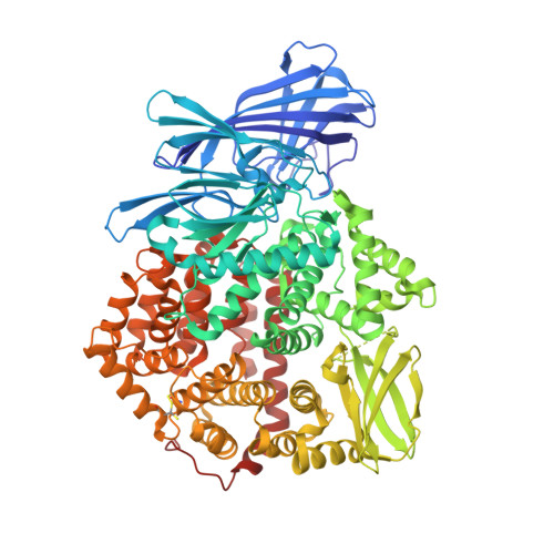
    Insulin-regulated aminopeptidase (IRAP) is an enzyme with several important biological functions that is known to process a large variety of different peptidic substrates, although the mechanism behind this wide specificity is not clearly understood. We describe a crystal structure of IRAP in complex with a recently developed bioactive and selective inhibitor at 2.53 Å resolution. In the presence of this inhibitor, the enzyme adopts a novel conformation in which domains II and IV are juxtaposed, forming a hollow structure that excludes external solvent access to the catalytic center. A loop adjacent to the enzyme's GAMEN motif undergoes structural reconfiguration, allowing the accommodation of bulky inhibitor side chains. Atomic interactions between the inhibitor and IRAP that are unique to this conformation can explain the strong selectivity compared to homologous aminopeptidases ERAP1 and ERAP2. This conformation provides insight on IRAP's catalytic cycle and reveals significant active-site plasticity that may underlie its substrate permissiveness.


  • Organizational Affiliation

    National Center for Scientific Research Demokritos, Agia Paraskevi , Athens 15310, Greece.


Macromolecules
Find similar proteins by:  (by identity cutoff)  |  3D Structure
Entity ID: 1
MoleculeChains Sequence LengthOrganismDetailsImage
Leucyl-cystinyl aminopeptidase
A, B
881Homo sapiensMutation(s): 0 
Gene Names: LNPEPOTASE
EC: 3.4.11.3
Membrane Entity: Yes 
UniProt & NIH Common Fund Data Resources
Find proteins for Q9UIQ6 (Homo sapiens)
Explore Q9UIQ6 
Go to UniProtKB:  Q9UIQ6
PHAROS:  Q9UIQ6
GTEx:  ENSG00000113441 
Entity Groups  
Sequence Clusters30% Identity50% Identity70% Identity90% Identity95% Identity100% Identity
UniProt GroupQ9UIQ6
Sequence Annotations
Expand
  • Reference Sequence
Oligosaccharides

Help

Entity ID: 2
MoleculeChains Length2D Diagram Glycosylation3D Interactions
alpha-D-mannopyranose-(1-3)-beta-D-mannopyranose-(1-4)-2-acetamido-2-deoxy-beta-D-glucopyranose-(1-4)-2-acetamido-2-deoxy-beta-D-glucopyranose
C, R
4N-Glycosylation
Glycosylation Resources
GlyTouCan:  G81315DD
GlyCosmos:  G81315DD
GlyGen:  G81315DD
Entity ID: 3
MoleculeChains Length2D Diagram Glycosylation3D Interactions
beta-D-mannopyranose-(1-4)-2-acetamido-2-deoxy-beta-D-glucopyranose-(1-4)-2-acetamido-2-deoxy-beta-D-glucopyranose
D, F, K, N, P
3N-Glycosylation
Glycosylation Resources
GlyTouCan:  G15407YE
GlyCosmos:  G15407YE
GlyGen:  G15407YE
Entity ID: 4
MoleculeChains Length2D Diagram Glycosylation3D Interactions
2-acetamido-2-deoxy-beta-D-glucopyranose-(1-4)-2-acetamido-2-deoxy-beta-D-glucopyranose
E, H, J, L, M
E, H, J, L, M, O, Q
2N-Glycosylation
Glycosylation Resources
GlyTouCan:  G42666HT
GlyCosmos:  G42666HT
GlyGen:  G42666HT
Entity ID: 5
MoleculeChains Length2D Diagram Glycosylation3D Interactions
alpha-D-mannopyranose-(1-3)-[alpha-D-mannopyranose-(1-6)]beta-D-mannopyranose-(1-4)-2-acetamido-2-deoxy-beta-D-glucopyranose-(1-4)-2-acetamido-2-deoxy-beta-D-glucopyranose
G, I
5N-Glycosylation
Glycosylation Resources
GlyTouCan:  G22768VO
GlyCosmos:  G22768VO
GlyGen:  G22768VO
Small Molecules
Ligands 4 Unique
IDChains Name / Formula / InChI Key2D Diagram3D Interactions
7O2
Query on 7O2

Download Ideal Coordinates CCD File 
X [auth A],
XA [auth B]
[(2~{S})-2-[[(2~{S})-1-azanyl-1-oxidanylidene-3-phenyl-propan-2-yl]carbamoyl]-4,4-diphenyl-butyl]-[(1~{R})-1-azanyl-3-phenyl-propyl]phosphinic acid
C35 H40 N3 O4 P
FDAGDZVCWKCEEX-NGYIUDBNSA-N
NAG
Query on NAG

Download Ideal Coordinates CCD File 
OA [auth B]
PA [auth B]
QA [auth B]
RA [auth B]
S [auth A]
OA [auth B],
PA [auth B],
QA [auth B],
RA [auth B],
S [auth A],
SA [auth B],
T [auth A],
TA [auth B],
U [auth A],
UA [auth B],
V [auth A],
VA [auth B]
2-acetamido-2-deoxy-beta-D-glucopyranose
C8 H15 N O6
OVRNDRQMDRJTHS-FMDGEEDCSA-N
BR
Query on BR

Download Ideal Coordinates CCD File 
AA [auth A]
AB [auth B]
BA [auth A]
BB [auth B]
CA [auth A]
AA [auth A],
AB [auth B],
BA [auth A],
BB [auth B],
CA [auth A],
CB [auth B],
DA [auth A],
EA [auth A],
FA [auth A],
GA [auth A],
HA [auth A],
IA [auth A],
JA [auth A],
KA [auth A],
LA [auth A],
MA [auth A],
NA [auth B],
Y [auth A],
YA [auth B],
Z [auth A],
ZA [auth B]
BROMIDE ION
Br
CPELXLSAUQHCOX-UHFFFAOYSA-M
ZN
Query on ZN

Download Ideal Coordinates CCD File 
W [auth A],
WA [auth B]
ZINC ION
Zn
PTFCDOFLOPIGGS-UHFFFAOYSA-N
Binding Affinity Annotations 
IDSourceBinding Affinity
7O2 BindingDB:  5MJ6 Ki: 18 (nM) from 1 assay(s)
IC50: 32 (nM) from 1 assay(s)
Experimental Data & Validation

Experimental Data

  • Method: X-RAY DIFFRACTION
  • Resolution: 2.53 Å
  • R-Value Free: 0.229 
  • R-Value Work: 0.174 
  • R-Value Observed: 0.177 
  • Space Group: P 21 21 21
Unit Cell:
Length ( Å )Angle ( ˚ )
a = 112.24α = 90
b = 143.17β = 90
c = 148.99γ = 90
Software Package:
Software NamePurpose
PHENIXrefinement
xia2data reduction
xia2data scaling
PHASERphasing

Structure Validation

View Full Validation Report


Ligand Structure Quality Assessment 


Entry History & Funding Information

Deposition Data


Funding OrganizationLocationGrant Number
European Community's Seventh Framework Programme (FP7/2007-2013)United KingdomBioStruct-X (grant agreement No 283570)
Education and Lifelong Learning? of the National Strategic Reference Framework: Research Funding Program of the General Secretariat for Research & TechnologyGreecegrant no. ERC-14 to ES
Harry J. Lloyd Charitable trustUnited States--

Revision History  (Full details and data files)

  • Version 1.0: 2017-04-05
    Type: Initial release
  • Version 1.1: 2017-04-26
    Changes: Database references
  • Version 2.0: 2020-07-29
    Type: Remediation
    Reason: Carbohydrate remediation
    Changes: Advisory, Atomic model, Data collection, Derived calculations, Structure summary
  • Version 2.1: 2024-01-17
    Changes: Data collection, Database references, Derived calculations, Refinement description, Structure summary