5C4I

Structure of an Oxalate Oxidoreductase


Experimental Data Snapshot

  • Method: X-RAY DIFFRACTION
  • Resolution: 2.27 Å
  • R-Value Free: 0.216 
  • R-Value Work: 0.174 
  • R-Value Observed: 0.176 

wwPDB Validation   3D Report Full Report


Ligand Structure Quality Assessment 


This is version 1.5 of the entry. See complete history


Literature

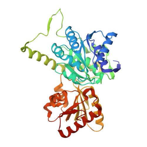
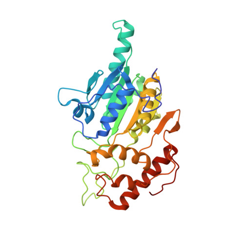
The Structure of an Oxalate Oxidoreductase Provides Insight into Microbial 2-Oxoacid Metabolism.

Gibson, M.I.Brignole, E.J.Pierce, E.Can, M.Ragsdale, S.W.Drennan, C.L.

(2015) Biochemistry 54: 4112-4120

  • DOI: https://doi.org/10.1021/acs.biochem.5b00521
  • Primary Citation of Related Structures:  
    5C4I

  • PubMed Abstract: 

    Thiamine pyrophosphate (TPP), a derivative of vitamin B1, is a versatile and ubiquitous cofactor. When coupled with [4Fe-4S] clusters in microbial 2-oxoacid:ferredoxin oxidoreductases (OFORs), TPP is involved in catalyzing low-potential redox reactions that are important for the synthesis of key metabolites and the reduction of N2, H(+), and CO2. We have determined the high-resolution (2.27 Å) crystal structure of the TPP-dependent oxalate oxidoreductase (OOR), an enzyme that allows microbes to grow on oxalate, a widely occurring dicarboxylic acid that is found in soil and freshwater and is responsible for kidney stone disease in humans. OOR catalyzes the anaerobic oxidation of oxalate, harvesting the low-potential electrons for use in anaerobic reduction and fixation of CO2. We compare the OOR structure to that of the only other structurally characterized OFOR family member, pyruvate:ferredoxin oxidoreductase. This side-by-side structural analysis highlights the key similarities and differences that are relevant for the chemistry of this entire class of TPP-utilizing enzymes.


  • Organizational Affiliation

    †Department of Chemistry, Massachusetts Institute of Technology, 77 Massachusetts Avenue, Cambridge, Massachusetts 02139, United States.


Macromolecules
Find similar proteins by:  (by identity cutoff)  |  3D Structure
Entity ID: 1
MoleculeChains Sequence LengthOrganismDetailsImage
Oxalate oxidoreductase subunit alpha
A, D
395Moorella thermoacetica ATCC 39073Mutation(s): 0 
EC: 1.2.7.10
UniProt
Find proteins for Q2RI41 (Moorella thermoacetica (strain ATCC 39073 / JCM 9320))
Explore Q2RI41 
Go to UniProtKB:  Q2RI41
Entity Groups  
Sequence Clusters30% Identity50% Identity70% Identity90% Identity95% Identity100% Identity
UniProt GroupQ2RI41
Sequence Annotations
Expand
  • Reference Sequence
Find similar proteins by:  (by identity cutoff)  |  3D Structure
Entity ID: 2
MoleculeChains Sequence LengthOrganismDetailsImage
Oxalate oxidoreductase subunit delta
B, E
315Moorella thermoacetica ATCC 39073Mutation(s): 0 
EC: 1.2.7.10
UniProt
Find proteins for Q2RI40 (Moorella thermoacetica (strain ATCC 39073 / JCM 9320))
Explore Q2RI40 
Go to UniProtKB:  Q2RI40
Entity Groups  
Sequence Clusters30% Identity50% Identity70% Identity90% Identity95% Identity100% Identity
UniProt GroupQ2RI40
Sequence Annotations
Expand
  • Reference Sequence
Find similar proteins by:  (by identity cutoff)  |  3D Structure
Entity ID: 3
MoleculeChains Sequence LengthOrganismDetailsImage
Oxalate oxidoreductase subunit beta
C, F
314Moorella thermoacetica ATCC 39073Mutation(s): 0 
EC: 1.2.7.10
UniProt
Find proteins for Q2RI42 (Moorella thermoacetica (strain ATCC 39073 / JCM 9320))
Explore Q2RI42 
Go to UniProtKB:  Q2RI42
Entity Groups  
Sequence Clusters30% Identity50% Identity70% Identity90% Identity95% Identity100% Identity
UniProt GroupQ2RI42
Sequence Annotations
Expand
  • Reference Sequence
Small Molecules
Ligands 4 Unique
IDChains Name / Formula / InChI Key2D Diagram3D Interactions
TPP
Query on TPP

Download Ideal Coordinates CCD File 
K [auth C],
P [auth F]
THIAMINE DIPHOSPHATE
C12 H19 N4 O7 P2 S
AYEKOFBPNLCAJY-UHFFFAOYSA-O
SF4
Query on SF4

Download Ideal Coordinates CCD File 
G [auth B]
H [auth B]
J [auth C]
M [auth E]
N [auth E]
G [auth B],
H [auth B],
J [auth C],
M [auth E],
N [auth E],
O [auth F]
IRON/SULFUR CLUSTER
Fe4 S4
LJBDFODJNLIPKO-UHFFFAOYSA-N
MG
Query on MG

Download Ideal Coordinates CCD File 
L [auth C],
Q [auth F]
MAGNESIUM ION
Mg
JLVVSXFLKOJNIY-UHFFFAOYSA-N
NA
Query on NA

Download Ideal Coordinates CCD File 
I [auth B],
R [auth F]
SODIUM ION
Na
FKNQFGJONOIPTF-UHFFFAOYSA-N
Experimental Data & Validation

Experimental Data

  • Method: X-RAY DIFFRACTION
  • Resolution: 2.27 Å
  • R-Value Free: 0.216 
  • R-Value Work: 0.174 
  • R-Value Observed: 0.176 
  • Space Group: P 21 21 21
Unit Cell:
Length ( Å )Angle ( ˚ )
a = 84.351α = 90
b = 152.203β = 90
c = 171.824γ = 90
Software Package:
Software NamePurpose
PHENIXrefinement
SCALEPACKdata scaling
PDB_EXTRACTdata extraction
PHASERphasing

Structure Validation

View Full Validation Report


Ligand Structure Quality Assessment 


Entry History & Funding Information

Deposition Data


Funding OrganizationLocationGrant Number
National Institutes of Health/National Institute of General Medical Sciences (NIH/NIGMS)United StatesGM069857
National Institutes of Health/National Institute of General Medical Sciences (NIH/NIGMS)United StatesGM39451
National Science Foundation (NSF, United States)United States1122374
Howard Hughes Medical Institute (HHMI)United States--
Martin 592 Family Society of Fellows for SustainabilityUnited States--

Revision History  (Full details and data files)

  • Version 1.0: 2015-07-01
    Type: Initial release
  • Version 1.1: 2015-07-22
    Changes: Database references
  • Version 1.2: 2016-03-16
    Changes: Data collection
  • Version 1.3: 2017-09-20
    Changes: Author supporting evidence, Derived calculations, Refinement description
  • Version 1.4: 2019-11-20
    Changes: Author supporting evidence
  • Version 1.5: 2024-03-06
    Changes: Data collection, Database references, Derived calculations