4ZB1

Crystal Structure of Blue Chromoprotein sgBP from Stichodactyla Gigantea


Experimental Data Snapshot

  • Method: X-RAY DIFFRACTION
  • Resolution: 2.25 Å
  • R-Value Free: 0.217 
  • R-Value Work: 0.174 
  • R-Value Observed: 0.176 

wwPDB Validation   3D Report Full Report


Ligand Structure Quality Assessment 


This is version 2.0 of the entry. See complete history


Literature

Chromophore Deprotonation State Alters the Optical Properties of Blue Chromoprotein

Chiang, C.Y.Lee, C.C.Lo, S.Y.Wang, A.H.Tsai, H.J.

(2015) PLoS One 10: e0134108-e0134108

  • DOI: https://doi.org/10.1371/journal.pone.0134108
  • Primary Citation of Related Structures:  
    4ZB1

  • PubMed Abstract: 

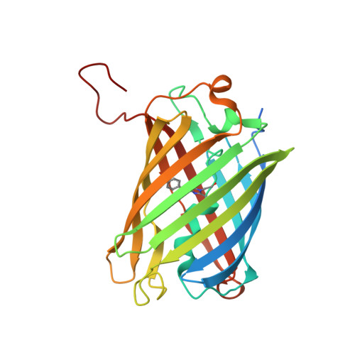
    Chromoproteins (CPs) have unique colors and can be used in biological applications. In this work, a novel blue CP with a maximum absorption peak (λmax) at 608 nm was identified from the carpet anemone Stichodactyla gigantea (sgBP). In vivo expression of sgBP in zebrafish would change the appearance of the fishes to have a blue color, indicating the potential biomarker function. To enhance the color properties, the crystal structure of sgBP at 2.25 Å resolution was determined to allow structure-based protein engineering. Among the mutations conducted in the Gln-Tyr-Gly chromophore and chromophore environment, a S157C mutation shifted the λmax to 604 nm with an extinction coefficient (ε) of 58,029 M-1·cm-1 and darkened the blue color expression. The S157C mutation in the sgBP chromophore environment could affect the color expression by altering the deprotonation state of the phenolic group in the chromophore. Our results provide a structural basis for the blue color enhancement of the biomarker development.


  • Organizational Affiliation

    Institute of Molecular and Cellular Biology, National Taiwan University, Taipei, Taiwan.


Macromolecules
Find similar proteins by:  (by identity cutoff)  |  3D Structure
Entity ID: 1
MoleculeChains Sequence LengthOrganismDetailsImage
Blue chromoprotein, sgBP
A, B
249Stichodactyla giganteaMutation(s): 0 
UniProt
Find proteins for A0A0M3KL34 (Stichodactyla gigantea)
Explore A0A0M3KL34 
Go to UniProtKB:  A0A0M3KL34
Entity Groups  
Sequence Clusters30% Identity50% Identity70% Identity90% Identity95% Identity100% Identity
UniProt GroupA0A0M3KL34
Sequence Annotations
Expand
  • Reference Sequence
Small Molecules
Ligands 1 Unique
IDChains Name / Formula / InChI Key2D Diagram3D Interactions
TOE
Query on TOE

Download Ideal Coordinates CCD File 
C [auth A],
D [auth B]
2-[2-(2-METHOXY-ETHOXY)-ETHOXY]-ETHOXYL
C7 H16 O4
JLGLQAWTXXGVEM-UHFFFAOYSA-N
Modified Residues  1 Unique
IDChains TypeFormula2D DiagramParent
CRQ
Query on CRQ
A, B
L-PEPTIDE LINKINGC16 H16 N4 O5GLN, TYR, GLY
Experimental Data & Validation

Experimental Data

  • Method: X-RAY DIFFRACTION
  • Resolution: 2.25 Å
  • R-Value Free: 0.217 
  • R-Value Work: 0.174 
  • R-Value Observed: 0.176 
  • Space Group: P 21 21 21
Unit Cell:
Length ( Å )Angle ( ˚ )
a = 61.095α = 90
b = 64.772β = 90
c = 154.736γ = 90
Software Package:
Software NamePurpose
REFMACrefinement
HKL-2000data reduction
MOLREPphasing
HKL-2000data scaling

Structure Validation

View Full Validation Report


Ligand Structure Quality Assessment 


Entry History 

Deposition Data

Revision History  (Full details and data files)

  • Version 1.0: 2015-08-12
    Type: Initial release
  • Version 1.1: 2023-11-08
    Changes: Data collection, Database references, Derived calculations, Refinement description
  • Version 2.0: 2023-11-15
    Changes: Atomic model, Data collection, Derived calculations