4XL8

Crystal Structure of Human Adenovirus 52 Short Fiber Knob in Complex with 2-O-Methyl-5-N-Acetylneuraminic Acid


Experimental Data Snapshot

  • Method: X-RAY DIFFRACTION
  • Resolution: 1.65 Å
  • R-Value Free: 0.174 
  • R-Value Work: 0.151 
  • R-Value Observed: 0.152 

wwPDB Validation   3D Report Full Report


Ligand Structure Quality Assessment 


This is version 2.3 of the entry. See complete history


Literature

Human Adenovirus 52 Uses Sialic Acid-containing Glycoproteins and the Coxsackie and Adenovirus Receptor for Binding to Target Cells.

Lenman, A.Liaci, A.M.Liu, Y.Ardahl, C.Rajan, A.Nilsson, E.Bradford, W.Kaeshammer, L.Jones, M.S.Frangsmyr, L.Feizi, T.Stehle, T.Arnberg, N.

(2015) PLoS Pathog 11: e1004657-e1004657

  • DOI: https://doi.org/10.1371/journal.ppat.1004657
  • Primary Citation of Related Structures:  
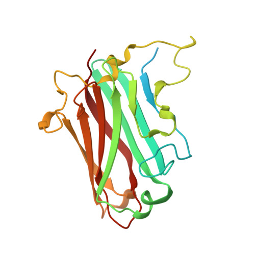
    4XL8

  • PubMed Abstract: 

    Most adenoviruses attach to host cells by means of the protruding fiber protein that binds to host cells via the coxsackievirus and adenovirus receptor (CAR) protein. Human adenovirus type 52 (HAdV-52) is one of only three gastroenteritis-causing HAdVs that are equipped with two different fiber proteins, one long and one short. Here we show, by means of virion-cell binding and infection experiments, that HAdV-52 can also attach to host cells via CAR, but most of the binding depends on sialylated glycoproteins. Glycan microarray, flow cytometry, surface plasmon resonance and ELISA analyses reveal that the terminal knob domain of the long fiber (52LFK) binds to CAR, and the knob domain of the short fiber (52SFK) binds to sialylated glycoproteins. X-ray crystallographic analysis of 52SFK in complex with 2-O-methylated sialic acid combined with functional studies of knob mutants revealed a new sialic acid binding site compared to other, known adenovirus:glycan interactions. Our findings shed light on adenovirus biology and may help to improve targeting of adenovirus-based vectors for gene therapy.


  • Organizational Affiliation

    Division of Virology, Department of Clinical Microbiology, Umeå University, Umeå, Sweden.


Macromolecules
Find similar proteins by:  (by identity cutoff)  |  3D Structure
Entity ID: 1
MoleculeChains Sequence LengthOrganismDetailsImage
Fiber-1
A, B, C
209human adenovirus 52Mutation(s): 0 
UniProt
Find proteins for A0MK70 (Human adenovirus 52)
Explore A0MK70 
Go to UniProtKB:  A0MK70
Entity Groups  
Sequence Clusters30% Identity50% Identity70% Identity90% Identity95% Identity100% Identity
UniProt GroupA0MK70
Sequence Annotations
Expand
  • Reference Sequence
Small Molecules
Ligands 7 Unique
IDChains Name / Formula / InChI Key2D Diagram3D Interactions
MNA
Query on MNA

Download Ideal Coordinates CCD File 
D [auth A],
I [auth C]
2-O-methyl-5-N-acetyl-alpha-D-neuraminic acid
C12 H21 N O9
NJRVVFURCKKXOD-MIDKXNQYSA-N
TAM
Query on TAM

Download Ideal Coordinates CCD File 
F [auth B]TRIS(HYDROXYETHYL)AMINOMETHANE
C7 H17 N O3
GKODZWOPPOTFGA-UHFFFAOYSA-N
GLU
Query on GLU

Download Ideal Coordinates CCD File 
G [auth B]GLUTAMIC ACID
C5 H9 N O4
WHUUTDBJXJRKMK-VKHMYHEASA-N
MRD
Query on MRD

Download Ideal Coordinates CCD File 
K [auth C](4R)-2-METHYLPENTANE-2,4-DIOL
C6 H14 O2
SVTBMSDMJJWYQN-RXMQYKEDSA-N
MPD
Query on MPD

Download Ideal Coordinates CCD File 
J [auth C](4S)-2-METHYL-2,4-PENTANEDIOL
C6 H14 O2
SVTBMSDMJJWYQN-YFKPBYRVSA-N
GOL
Query on GOL

Download Ideal Coordinates CCD File 
E [auth A]GLYCEROL
C3 H8 O3
PEDCQBHIVMGVHV-UHFFFAOYSA-N
ALA
Query on ALA

Download Ideal Coordinates CCD File 
H [auth B]ALANINE
C3 H7 N O2
QNAYBMKLOCPYGJ-REOHCLBHSA-N
Experimental Data & Validation

Experimental Data

  • Method: X-RAY DIFFRACTION
  • Resolution: 1.65 Å
  • R-Value Free: 0.174 
  • R-Value Work: 0.151 
  • R-Value Observed: 0.152 
  • Space Group: P 21 21 21
Unit Cell:
Length ( Å )Angle ( ˚ )
a = 63.77α = 90
b = 81.61β = 90
c = 92.65γ = 90
Software Package:
Software NamePurpose
PHENIXrefinement
PDB_EXTRACTdata extraction
XDSdata reduction
XSCALEdata scaling
PHASERphasing

Structure Validation

View Full Validation Report


Ligand Structure Quality Assessment 


Entry History & Funding Information

Deposition Data


Funding OrganizationLocationGrant Number
German Research FoundationGermanySFB 685

Revision History  (Full details and data files)

  • Version 1.0: 2015-01-21
    Type: Initial release
  • Version 1.1: 2015-02-11
    Changes: Other
  • Version 1.2: 2015-03-04
    Changes: Database references
  • Version 1.3: 2015-07-22
    Changes: Database references
  • Version 2.0: 2017-09-06
    Changes: Advisory, Atomic model, Author supporting evidence, Derived calculations
  • Version 2.1: 2019-10-16
    Changes: Data collection
  • Version 2.2: 2020-07-29
    Type: Remediation
    Reason: Carbohydrate remediation
    Changes: Data collection, Derived calculations, Structure summary
  • Version 2.3: 2024-01-10
    Changes: Data collection, Database references, Refinement description, Structure summary