


Crystal Structure of Frutalin from Artocarpus incisa
Pereira, H.M., Moreira, A.C.O.M., Vieira Neto, A.E., Moreno, F.B.M.B., Lobo, M.D.P., Sousa, F.D., Grangeiro, T.B., Moreira, R.A.To be published.
Experimental Data Snapshot
wwPDB Validation 3D Report Full Report

Entity ID: 1 | |||||
|---|---|---|---|---|---|
| Molecule | Chains | Sequence Length | Organism | Details | Image |
| Frutalin | 157 | Artocarpus | Mutation(s): 0 | 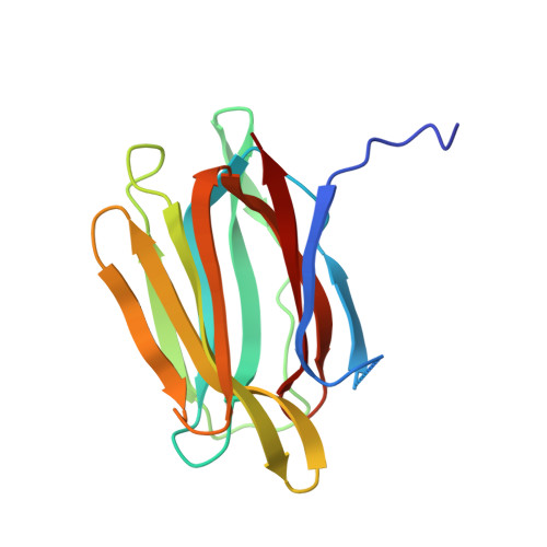 | |
UniProt | |||||
Find proteins for A0A0R4I968 (Artocarpus) Explore A0A0R4I968 Go to UniProtKB: A0A0R4I968 | |||||
Entity Groups | |||||
| Sequence Clusters | 30% Identity50% Identity70% Identity90% Identity95% Identity100% Identity | ||||
| UniProt Group | A0A0R4I968 | ||||
Sequence AnnotationsExpand | |||||
| |||||
| Length ( Å ) | Angle ( ˚ ) |
|---|---|
| a = 76.17 | α = 90 |
| b = 74.36 | β = 96.57 |
| c = 118.986 | γ = 90 |
| Software Name | Purpose |
|---|---|
| d*TREK | data scaling |
| Aimless | data scaling |
| PHASER | phasing |
| PHENIX | refinement |
| PDB_EXTRACT | data extraction |
RCSB PDB (citation) is hosted by
RCSB PDB is a member of the

