4UTG

Burkholderia pseudomallei heptokinase WcbL,AMPPNP (ATP analogue) complex.


Experimental Data Snapshot

  • Method: X-RAY DIFFRACTION
  • Resolution: 1.93 Å
  • R-Value Free: 0.242 
  • R-Value Work: 0.194 
  • R-Value Observed: 0.196 

wwPDB Validation   3D Report Full Report


Ligand Structure Quality Assessment 


This is version 1.1 of the entry. See complete history


Literature

Unraveling the B.Pseudomallei Heptokinase Wcbl: From Structure to Drug Discovery.

Vivoli, M.Isupov, M.N.Nicholas, R.Hill, A.Scott, A.E.Kosma, P.Prior, J.L.Harmer, N.J.

(2015) Chem Biol 22: 1622

  • DOI: https://doi.org/10.1016/j.chembiol.2015.10.015
  • Primary Citation of Related Structures:  
    4USK, 4USM, 4UT4, 4UTG

  • PubMed Abstract: 

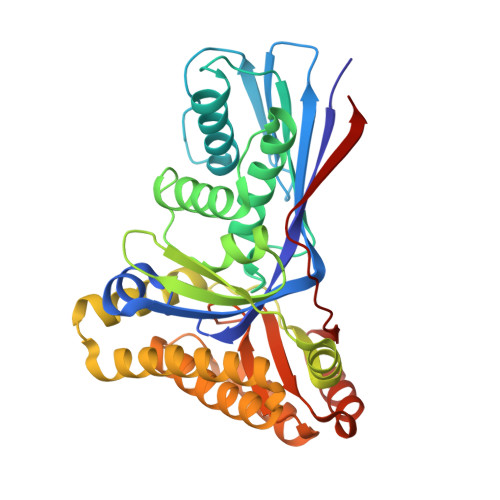
    Gram-negative bacteria utilize heptoses as part of their repertoire of extracellular polysaccharide virulence determinants. Disruption of heptose biosynthesis offers an attractive target for novel antimicrobials. A critical step in the synthesis of heptoses is their 1-O phosphorylation, mediated by kinases such as HldE or WcbL. Here, we present the structure of WcbL from Burkholderia pseudomallei. We report that WcbL operates through a sequential ordered Bi-Bi mechanism, loading the heptose first and then ATP. We show that dimeric WcbL binds ATP anti-cooperatively in the absence of heptose, and cooperatively in its presence. Modeling of WcbL suggests that heptose binding causes an elegant switch in the hydrogen-bonding network, facilitating the binding of a second ATP molecule. Finally, we screened a library of drug-like fragments, identifying hits that potently inhibit WcbL. Our results provide a novel mechanism for control of substrate binding and emphasize WcbL as an attractive anti-microbial target for Gram-negative bacteria.


  • Organizational Affiliation

    Department of Biosciences, University of Exeter, Henry Wellcome Building, Stocker Road, Exeter EX4 4QD, UK.


Macromolecules
Find similar proteins by:  (by identity cutoff)  |  3D Structure
Entity ID: 1
MoleculeChains Sequence LengthOrganismDetailsImage
SUGAR KINASE
A, B
346Burkholderia pseudomallei K96243Mutation(s): 3 
EC: 2.7.1.167
UniProt
Find proteins for H7C745 (Burkholderia pseudomallei (strain K96243))
Explore H7C745 
Go to UniProtKB:  H7C745
Entity Groups  
Sequence Clusters30% Identity50% Identity70% Identity90% Identity95% Identity100% Identity
UniProt GroupH7C745
Sequence Annotations
Expand
  • Reference Sequence
Small Molecules
Ligands 4 Unique
IDChains Name / Formula / InChI Key2D Diagram3D Interactions
ANP
Query on ANP

Download Ideal Coordinates CCD File 
C [auth A],
H [auth B]
PHOSPHOAMINOPHOSPHONIC ACID-ADENYLATE ESTER
C10 H17 N6 O12 P3
PVKSNHVPLWYQGJ-KQYNXXCUSA-N
EDO
Query on EDO

Download Ideal Coordinates CCD File 
G [auth A],
L [auth B]
1,2-ETHANEDIOL
C2 H6 O2
LYCAIKOWRPUZTN-UHFFFAOYSA-N
CL
Query on CL

Download Ideal Coordinates CCD File 
E [auth A],
F [auth A],
J [auth B],
K [auth B]
CHLORIDE ION
Cl
VEXZGXHMUGYJMC-UHFFFAOYSA-M
MG
Query on MG

Download Ideal Coordinates CCD File 
D [auth A],
I [auth B]
MAGNESIUM ION
Mg
JLVVSXFLKOJNIY-UHFFFAOYSA-N
Experimental Data & Validation

Experimental Data

  • Method: X-RAY DIFFRACTION
  • Resolution: 1.93 Å
  • R-Value Free: 0.242 
  • R-Value Work: 0.194 
  • R-Value Observed: 0.196 
  • Space Group: C 2 2 21
Unit Cell:
Length ( Å )Angle ( ˚ )
a = 101.44α = 90
b = 115.02β = 90
c = 166.78γ = 90
Software Package:
Software NamePurpose
xia2data reduction
XDSdata reduction
Aimlessdata scaling
SHELXphasing
ARP/wARPphasing
REFMACrefinement

Structure Validation

View Full Validation Report


Ligand Structure Quality Assessment 


Entry History 

Deposition Data

Revision History  (Full details and data files)

  • Version 1.0: 2016-01-13
    Type: Initial release
  • Version 1.1: 2017-06-28
    Changes: Refinement description