


A Structural Snapshot of Type II Pilus Formation in Streptococcus pneumoniae.
Shaik, M.M., Lombardi, C., Maragno Trindade, D., Fenel, D., Schoehn, G., Di Guilmi, A.M., Dessen, A.(2015) J Biol Chem 290: 22581-22592
- PubMed: 26198632
- DOI: https://doi.org/10.1074/jbc.M115.647834
- Primary Citation of Related Structures:
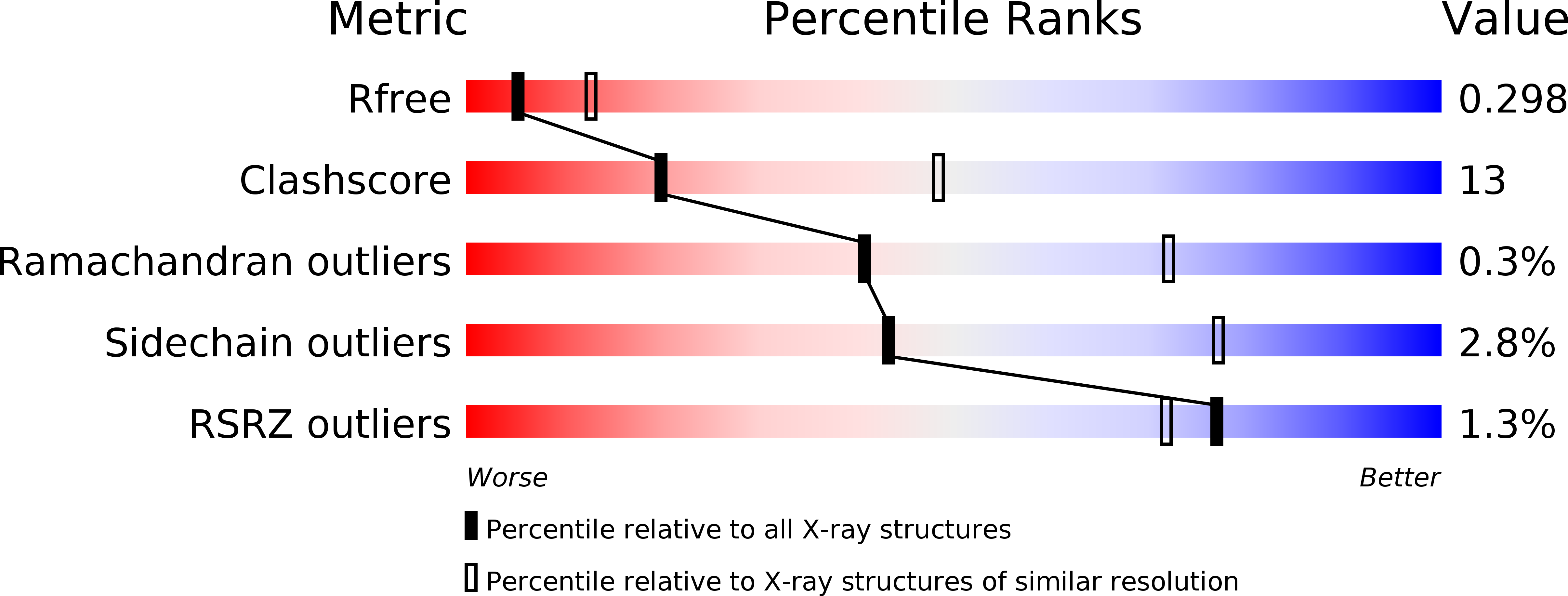
4S3L, 4Y4Q - PubMed Abstract:
Pili are fibrous appendages expressed on the surface of a vast number of bacterial species, and their role in surface adhesion is important for processes such as infection, colonization, andbiofilm formation. The human pathogen Streptococcus pneumoniae expresses two different types of pili, PI-1 and PI-2, both of which require the concerted action of structural proteins and sortases for their polymerization. The type PI-1 streptococcal pilus is a complex, well studied structure, but the PI-2 type, present in a number of invasive pneumococcal serotypes, has to date remained less well understood. The PI-2 pilus consists of repeated units of a single protein, PitB, whose covalent association is catalyzed by cognate sortase SrtG-1 and partner protein SipA. Here we report the high resolution crystal structures of PitB and SrtG1 and use molecular modeling to visualize a "trapped" 1:1 complex between the two molecules. X-ray crystallography and electron microscopy reveal that the pneumococcal PI-2 backbone fiber is formed by PitB monomers associated in head-to-tail fashion and that short, flexible fibers can be formed even in the absence of coadjuvant proteins. These observations, obtained with a simple pilus biosynthetic system, are likely to be applicable to other fiber formation processes in a variety of Gram-positive organisms.
Organizational Affiliation:
From the Université Grenoble Alpes, Institut de Biologie Structurale (IBS), F-38044 Grenoble, France, CNRS, IBS, 38044 Grenoble, France, Commissariat à l'Energie Atomique, IBS, Grenoble, France, and.


