4R1F

Re-refined Human DNA topoisomerase IIa (ATPase and transducer domains) in complex with ADP and SO4


Experimental Data Snapshot

  • Method: X-RAY DIFFRACTION
  • Resolution: 2.51 Å
  • R-Value Free: 0.243 
  • R-Value Work: 0.208 
  • R-Value Observed: 0.211 

wwPDB Validation   3D Report Full Report


Ligand Structure Quality Assessment 


This is version 1.2 of the entry. See complete history

Re-refinement Note

This entry reflects an alternative modeling of the original data in: 1ZXN


Literature

Structure of the N-Terminal Gyrase B Fragment in Complex with ADPPi Reveals Rigid-Body Motion Induced by ATP Hydrolysis

Stanger, F.V.Dehio, C.Schirmer, T.

(2014) PLoS One 9: e107289-e107289

  • DOI: https://doi.org/10.1371/journal.pone.0107289
  • Primary Citation of Related Structures:  
    4PRV, 4PRX, 4PU9, 4R1F

  • PubMed Abstract: 

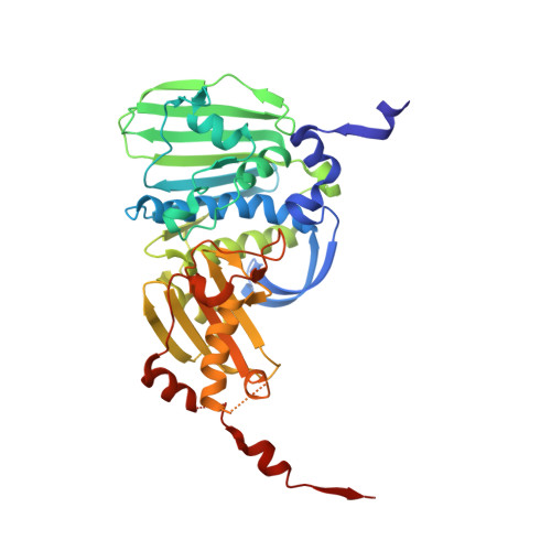
    Type II DNA topoisomerases are essential enzymes that catalyze topological rearrangement of double-stranded DNA using the free energy generated by ATP hydrolysis. Bacterial DNA gyrase is a prototype of this family and is composed of two subunits (GyrA, GyrB) that form a GyrA2GyrB2 heterotetramer. The N-terminal 43-kDa fragment of GyrB (GyrB43) from E. coli comprising the ATPase and the transducer domains has been studied extensively. The dimeric fragment is competent for ATP hydrolysis and its structure in complex with the substrate analog AMPPNP is known. Here, we have determined the remaining conformational states of the enzyme along the ATP hydrolysis reaction path by solving crystal structures of GyrB43 in complex with ADP⋅BeF3, ADP⋅Pi, and ADP. Upon hydrolysis, the enzyme undergoes an obligatory 12° domain rearrangement to accommodate the 1.5 Å increase in distance between the γ- and β-phosphate of the nucleotide within the sealed binding site at the domain interface. Conserved residues from the QTK loop of the transducer domain (also part of the domain interface) couple the small structural change within the binding site with the rigid body motion. The domain reorientation is reflected in a significant 7 Å increase in the separation of the two transducer domains of the dimer that would embrace one of the DNA segments in full-length gyrase. The observed conformational change is likely to be relevant for the allosteric coordination of ATP hydrolysis with DNA binding, cleavage/re-ligation and/or strand passage.


  • Organizational Affiliation

    Focal Area Structural Biology and Biophysics, Biozentrum, University of Basel, Basel, Switzerland; Focal Area Infection Biology, Biozentrum, University of Basel, Basel, Switzerland.


Macromolecules
Find similar proteins by:  (by identity cutoff)  |  3D Structure
Entity ID: 1
MoleculeChains Sequence LengthOrganismDetailsImage
DNA topoisomerase 2-alpha
A, B, C, D
400Homo sapiensMutation(s): 0 
Gene Names: TOP2TOP2A
EC: 5.99.1.3
UniProt & NIH Common Fund Data Resources
Find proteins for P11388 (Homo sapiens)
Explore P11388 
Go to UniProtKB:  P11388
PHAROS:  P11388
GTEx:  ENSG00000131747 
Entity Groups  
Sequence Clusters30% Identity50% Identity70% Identity90% Identity95% Identity100% Identity
UniProt GroupP11388
Sequence Annotations
Expand
  • Reference Sequence
Small Molecules
Ligands 4 Unique
IDChains Name / Formula / InChI Key2D Diagram3D Interactions
ADP
Query on ADP

Download Ideal Coordinates CCD File 
F [auth A],
J [auth B],
L [auth C],
P [auth D]
ADENOSINE-5'-DIPHOSPHATE
C10 H15 N5 O10 P2
XTWYTFMLZFPYCI-KQYNXXCUSA-N
SO4
Query on SO4

Download Ideal Coordinates CCD File 
I [auth B],
M [auth C],
O [auth D]
SULFATE ION
O4 S
QAOWNCQODCNURD-UHFFFAOYSA-L
GOL
Query on GOL

Download Ideal Coordinates CCD File 
G [auth A]GLYCEROL
C3 H8 O3
PEDCQBHIVMGVHV-UHFFFAOYSA-N
MG
Query on MG

Download Ideal Coordinates CCD File 
E [auth A],
H [auth B],
K [auth C],
N [auth D]
MAGNESIUM ION
Mg
JLVVSXFLKOJNIY-UHFFFAOYSA-N
Experimental Data & Validation

Experimental Data

  • Method: X-RAY DIFFRACTION
  • Resolution: 2.51 Å
  • R-Value Free: 0.243 
  • R-Value Work: 0.208 
  • R-Value Observed: 0.211 
  • Space Group: P 1 21 1
Unit Cell:
Length ( Å )Angle ( ˚ )
a = 71.015α = 90
b = 90.492β = 89.97
c = 148.293γ = 90
Software Package:
Software NamePurpose
REFMACrefinement
PDB_EXTRACTdata extraction

Structure Validation

View Full Validation Report


Ligand Structure Quality Assessment 


Entry History 

Deposition Data

Revision History  (Full details and data files)

  • Version 1.0: 2014-09-24
    Type: Initial release
  • Version 1.1: 2014-11-05
    Changes: Other
  • Version 1.2: 2023-11-08
    Changes: Data collection, Database references, Derived calculations, Refinement description