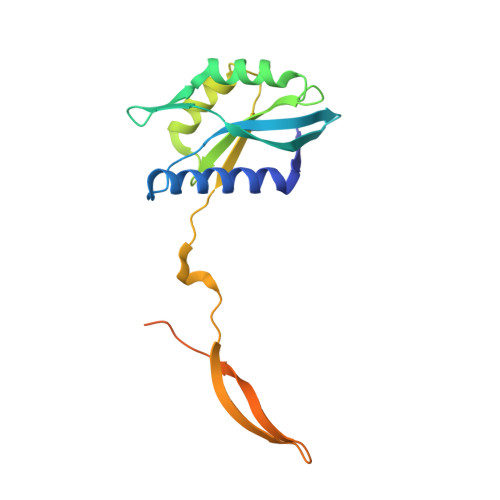
Crystal structure of the human CSN6 MPN domain
Ma, X.L., Xu, M., Jiang, T.(2014) Biochem Biophys Res Commun 453: 25-30
- PubMed: 25242525
- DOI: https://doi.org/10.1016/j.bbrc.2014.09.046
- Primary Citation of Related Structures:
4R14 - PubMed Abstract:
The mammalian COP9 signalosome is an eight-subunit (CSN1-CSN8) complex that plays essential roles in multiple cellular and physiological processes. CSN5 and CSN6 are the only two MPN (Mpr1-Pad1-N-terminal) domain-containing subunits in the complex. Unlike the CSN5 MPN domain, CSN6 lacks a metal-binding site and isopeptidase activity. Here, we report the crystal structure of the human CSN6 MPN domain. Each CSN6 monomer contains nine β sheets surrounded by three helices. Two forms of dimers are observed in the crystal structure. Interestingly, a domain swapping of β8 and β9 strands occurs between two neighboring monomers to complete a typical MPN fold. Analyses of the pseudo metal-binding motif in CSN6 suggest that the loss of two key histidine residues may contribute to the lack of catalytic activity in CSN6. Comparing the MPN domain of our CSN6 with that in the CSN complex shows that apart from the different β8-β9 conformation, they have minor conformational differences at two insertion regions (Ins-1 and Ins-2). Besides, the interacting mode of CSN6-CSN6 in our structure is distinct from that of CSN5-CSN6 in the CSN complex structure. Moreover, the functional implications for Ins-1 and Ins-2 are discussed.
Organizational Affiliation:
National Laboratory of Biomacromolecules, Institute of Biophysics, Chinese Academy of Sciences, Beijing 100101, China; University of Chinese Academy of Sciences, 19A Yuquan Road, Shijingshan District, Beijing 100039, China.