4QX3

Cry3A Toxin structure obtained by injecting Bacillus thuringiensis cells in an XFEL beam, collecting data by serial femtosecond crystallographic methods and processing data with the CrystFEL software suite


Experimental Data Snapshot

  • Method: X-RAY DIFFRACTION
  • Resolution: 2.90 Å
  • R-Value Free: 0.194 
  • R-Value Work: 0.177 
  • R-Value Observed: 0.178 

wwPDB Validation   3D Report Full Report


This is version 1.4 of the entry. See complete history


Literature

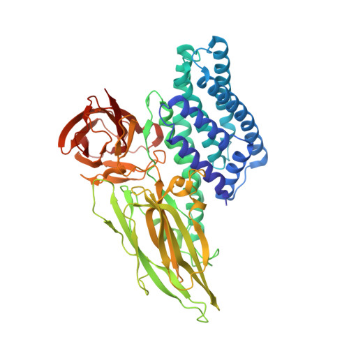
Protein crystal structure obtained at 2.9 angstrom resolution from injecting bacterial cells into an X-ray free-electron laser beam.

Sawaya, M.R.Cascio, D.Gingery, M.Rodriguez, J.Goldschmidt, L.Colletier, J.P.Messerschmidt, M.M.Boutet, S.Koglin, J.E.Williams, G.J.Brewster, A.S.Nass, K.Hattne, J.Botha, S.Doak, R.B.Shoeman, R.L.DePonte, D.P.Park, H.W.Federici, B.A.Sauter, N.K.Schlichting, I.Eisenberg, D.S.

(2014) Proc Natl Acad Sci U S A 111: 12769-12774

  • DOI: https://doi.org/10.1073/pnas.1413456111
  • Primary Citation of Related Structures:  
    4QX0, 4QX1, 4QX2, 4QX3

  • PubMed Abstract: 

    It has long been known that toxins produced by Bacillus thuringiensis (Bt) are stored in the bacterial cells in crystalline form. Here we describe the structure determination of the Cry3A toxin found naturally crystallized within Bt cells. When whole Bt cells were streamed into an X-ray free-electron laser beam we found that scattering from other cell components did not obscure diffraction from the crystals. The resolution limits of the best diffraction images collected from cells were the same as from isolated crystals. The integrity of the cells at the moment of diffraction is unclear; however, given the short time (∼ 5 µs) between exiting the injector to intersecting with the X-ray beam, our result is a 2.9-Å-resolution structure of a crystalline protein as it exists in a living cell. The study suggests that authentic in vivo diffraction studies can produce atomic-level structural information.


  • Organizational Affiliation

    UCLA-DOE Institute for Genomics and Proteomics, Department of Biological Chemistry, and.


Macromolecules
Find similar proteins by:  (by identity cutoff)  |  3D Structure
Entity ID: 1
MoleculeChains Sequence LengthOrganismDetailsImage
Pesticidal crystal protein cry3Aa587Bacillus thuringiensis serovar tenebrionisMutation(s): 0 
Gene Names: bt13cry3Acry3AacryCcryIIIacryIIIA(a)
UniProt
Find proteins for P0A379 (Bacillus thuringiensis subsp. tenebrionis)
Explore P0A379 
Go to UniProtKB:  P0A379
Entity Groups  
Sequence Clusters30% Identity50% Identity70% Identity90% Identity95% Identity100% Identity
UniProt GroupP0A379
Sequence Annotations
Expand
  • Reference Sequence
Experimental Data & Validation

Experimental Data

  • Method: X-RAY DIFFRACTION
  • Resolution: 2.90 Å
  • R-Value Free: 0.194 
  • R-Value Work: 0.177 
  • R-Value Observed: 0.178 
  • Space Group: C 2 2 21
Unit Cell:
Length ( Å )Angle ( ˚ )
a = 117.3α = 90
b = 135.3β = 90
c = 105.3γ = 90
Software Package:
Software NamePurpose
BUSTER-TNTrefinement
PDB_EXTRACTdata extraction
CrystFELdata reduction
CrystFELdata scaling
BUSTERrefinement

Structure Validation

View Full Validation Report


Entry History 

Revision History  (Full details and data files)

  • Version 1.0: 2014-08-13
    Type: Initial release
  • Version 1.1: 2014-09-03
    Changes: Database references
  • Version 1.2: 2014-09-24
    Changes: Database references
  • Version 1.3: 2018-02-14
    Changes: Data collection
  • Version 1.4: 2024-02-28
    Changes: Data collection, Database references