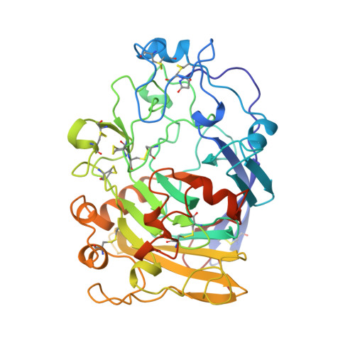
4OVW

ENDOGLUCANASE I COMPLEXED WITH EPOXYBUTYL CELLOBIOSE


Experimental Data Snapshot

  • Method: X-RAY DIFFRACTION
  • Resolution: 2.30 Å
  • R-Value Free: 0.270 
  • R-Value Work: 0.180 

wwPDB Validation   3D Report Full Report


This is version 3.1 of the entry. See complete history


Literature

Structure of the endoglucanase I from Fusarium oxysporum: native, cellobiose, and 3,4-epoxybutyl beta-D-cellobioside-inhibited forms, at 2.3 A resolution.

Sulzenbacher, G.Schulein, M.Davies, G.J.

(1997) Biochemistry 36: 5902-5911

  • DOI: https://doi.org/10.1021/bi962963+
  • Primary Citation of Related Structures:  
    2OVW, 3OVW, 4OVW

  • PubMed Abstract: 

    The mechanisms involved in the enzymatic degradation of cellulose are of great ecological and commercial importance. The breakdown of cellulose by fungal species is performed by a consortium of free enzymes, known as cellobiohydrolases and endoglucanases, which are found in many of the 57 glycosyl hydrolase families. The structure of the endoglucanase I (EG I), found in glycosyl hydrolase family 7, from the thermophilic fungus Fusarium oxysporum has been solved at 2.3 A resolution. In addition to the native enzyme, structures have also been determined with both the affinity label, 3,4-epoxybutyl beta-D-cellobioside, and the reaction product cellobiose. The affinity label is covalently bound, as expected, to the catalytic nucleophile, Glu197, with clear evidence for binding of both the R and S stereoisomers. Cellobiose is found bound to the -2 and -1 subsites of the enzyme. In marked contrast to the structure of EG I with a nonhydrolyzable thiosaccharide analog, which spanned the -2, -1, and +1 subsites and which had a skew-boat conformation for the -1 subsite sugar [Sulzenbacher, G., et al. (1996) Biochemistry 35, 15280-15287], the cellobiose complex shows no pyranoside ring distortion in the -1 subsite, implying that strain is induced primarily by the additional +1 subsite interactions and that the product is found, as expected, in its unstrained conformation.


  • Organizational Affiliation

    Department of Chemistry, University of York, Heslington, U.K.


Macromolecules
Find similar proteins by:  (by identity cutoff)  |  3D Structure
Entity ID: 1
MoleculeChains Sequence LengthOrganismDetailsImage
ENDOGLUCANASE I
A, B
411Fusarium oxysporumMutation(s): 0 
EC: 3.2.1.4
UniProt
Find proteins for P46237 (Fusarium oxysporum)
Explore P46237 
Go to UniProtKB:  P46237
Entity Groups  
Sequence Clusters30% Identity50% Identity70% Identity90% Identity95% Identity100% Identity
UniProt GroupP46237
Sequence Annotations
Expand
  • Reference Sequence
Small Molecules
Ligands 2 Unique
IDChains Name / Formula / InChI Key2D Diagram3D Interactions
IN1
Query on IN1

Download Ideal Coordinates CCD File 
D [auth A],
F [auth B]
4-(beta-D-glucopyranosyloxy)-2,2-dihydroxybutyl propanoate
C13 H24 O10
LNJDDDXXDSOXEJ-DVYMNCLGSA-N
NAG
Query on NAG

Download Ideal Coordinates CCD File 
C [auth A],
E [auth B]
2-acetamido-2-deoxy-beta-D-glucopyranose
C8 H15 N O6
OVRNDRQMDRJTHS-FMDGEEDCSA-N
Modified Residues  1 Unique
IDChains TypeFormula2D DiagramParent
PCA
Query on PCA
A, B
L-PEPTIDE LINKINGC5 H7 N O3GLN
Experimental Data & Validation

Experimental Data

  • Method: X-RAY DIFFRACTION
  • Resolution: 2.30 Å
  • R-Value Free: 0.270 
  • R-Value Work: 0.180 
  • Space Group: P 1 21 1
Unit Cell:
Length ( Å )Angle ( ˚ )
a = 65.94α = 90
b = 82.62β = 94.16
c = 73.16γ = 90
Software Package:
Software NamePurpose
DENZOdata reduction
SCALEPACKdata scaling
AMoREphasing
REFMACrefinement

Structure Validation

View Full Validation Report


Entry History 

Deposition Data

Revision History  (Full details and data files)

  • Version 1.0: 1998-04-08
    Type: Initial release
  • Version 1.1: 2008-03-03
    Changes: Version format compliance
  • Version 1.2: 2011-07-13
    Changes: Non-polymer description, Version format compliance
  • Version 2.0: 2019-12-25
    Changes: Advisory, Derived calculations, Polymer sequence
  • Version 3.0: 2020-07-29
    Type: Remediation
    Reason: Carbohydrate remediation
    Changes: Atomic model, Data collection, Derived calculations, Structure summary
  • Version 3.1: 2024-04-03
    Changes: Data collection, Database references, Refinement description, Structure summary