4NTD

Crystal structure of HlmI


Experimental Data Snapshot

  • Method: X-RAY DIFFRACTION
  • Resolution: 1.60 Å
  • R-Value Free: 0.159 
  • R-Value Work: 0.146 
  • R-Value Observed: 0.147 

wwPDB Validation   3D Report Full Report


Ligand Structure Quality Assessment 


This is version 1.1 of the entry. See complete history


Literature

Flavoenzyme-Catalyzed Formation of Disulfide Bonds in Natural Products

Scharf, D.H.Groll, M.Habel, A.Heinekamp, T.Hertweck, C.Brakhage, A.A.Huber, E.M.

(2014) Angew Chem Int Ed Engl 53: 2221-2224

  • DOI: https://doi.org/10.1002/anie.201309302
  • Primary Citation of Related Structures:  
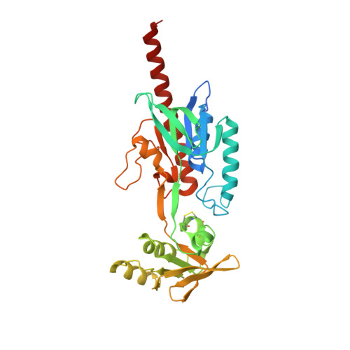
    4NTC, 4NTD, 4NTE

  • PubMed Abstract: 

    Nature provides a rich source of compounds with diverse chemical structures and biological activities, among them, sulfur-containing metabolites from bacteria and fungi. Some of these compounds bear a disulfide moiety that is indispensable for their bioactivity. Specialized oxidoreductases such as GliT, HlmI, and DepH catalyze the formation of this disulfide bridge in the virulence factor gliotoxin, the antibiotic holomycin, and the anticancer drug romidepsin, respectively. We have examined all three enzymes by X-ray crystallography and activity assays. Despite their differently sized substrate binding clefts and hence, their diverse substrate preferences, a unifying reaction mechanism is proposed based on the obtained crystal structures and further supported by mutagenesis experiments.


  • Organizational Affiliation

    Departments of Molecular and Applied Microbiology and Biomolecular Chemistry, Leibniz Institute for Natural Product Research and Infection Biology, HKI, and Friedrich Schiller University Jena, Beutenbergstrasse 11a, 07745 Jena (Germany).


Macromolecules
Find similar proteins by:  (by identity cutoff)  |  3D Structure
Entity ID: 1
MoleculeChains Sequence LengthOrganismDetailsImage
Thioredoxin reductase340Streptomyces clavuligerusMutation(s): 0 
Gene Names: SCLAV_5275
UniProt
Find proteins for E2PZ87 (Streptomyces clavuligerus)
Explore E2PZ87 
Go to UniProtKB:  E2PZ87
Entity Groups  
Sequence Clusters30% Identity50% Identity70% Identity90% Identity95% Identity100% Identity
UniProt GroupE2PZ87
Sequence Annotations
Expand
  • Reference Sequence
Small Molecules
Ligands 5 Unique
IDChains Name / Formula / InChI Key2D Diagram3D Interactions
FAD
Query on FAD

Download Ideal Coordinates CCD File 
C [auth A]FLAVIN-ADENINE DINUCLEOTIDE
C27 H33 N9 O15 P2
VWWQXMAJTJZDQX-UYBVJOGSSA-N
GSH
Query on GSH

Download Ideal Coordinates CCD File 
B [auth A]GLUTATHIONE
C10 H17 N3 O6 S
RWSXRVCMGQZWBV-WDSKDSINSA-N
PO4
Query on PO4

Download Ideal Coordinates CCD File 
F [auth A]PHOSPHATE ION
O4 P
NBIIXXVUZAFLBC-UHFFFAOYSA-K
GOL
Query on GOL

Download Ideal Coordinates CCD File 
D [auth A]GLYCEROL
C3 H8 O3
PEDCQBHIVMGVHV-UHFFFAOYSA-N
CL
Query on CL

Download Ideal Coordinates CCD File 
E [auth A]CHLORIDE ION
Cl
VEXZGXHMUGYJMC-UHFFFAOYSA-M
Experimental Data & Validation

Experimental Data

  • Method: X-RAY DIFFRACTION
  • Resolution: 1.60 Å
  • R-Value Free: 0.159 
  • R-Value Work: 0.146 
  • R-Value Observed: 0.147 
  • Space Group: P 31 2 1
Unit Cell:
Length ( Å )Angle ( ˚ )
a = 96.35α = 90
b = 96.35β = 90
c = 133.49γ = 120
Software Package:
Software NamePurpose
XDSdata scaling
PHASERphasing
REFMACrefinement
XDSdata reduction
XSCALEdata scaling

Structure Validation

View Full Validation Report


Ligand Structure Quality Assessment 


Entry History 

Deposition Data

Revision History  (Full details and data files)

  • Version 1.0: 2014-03-05
    Type: Initial release
  • Version 1.1: 2023-11-08
    Changes: Data collection, Database references, Derived calculations, Refinement description