4JUO

A low-resolution three-gate structure of topoisomerase IV from Streptococcus pneumoniae in space group H32


Experimental Data Snapshot

  • Method: X-RAY DIFFRACTION
  • Resolution: 6.53 Å
  • R-Value Free: 0.298 
  • R-Value Work: 0.247 
  • R-Value Observed: 0.250 

wwPDB Validation   3D Report Full Report


This is version 1.4 of the entry. See complete history


Literature

Structure of an 'open' clamp type II topoisomerase-DNA complex provides a mechanism for DNA capture and transport.

Laponogov, I.Veselkov, D.A.Crevel, I.M.Pan, X.S.Fisher, L.M.Sanderson, M.R.

(2013) Nucleic Acids Res 41: 9911-9923

  • DOI: https://doi.org/10.1093/nar/gkt749
  • Primary Citation of Related Structures:  
    4I3H, 4JUO

  • PubMed Abstract: 

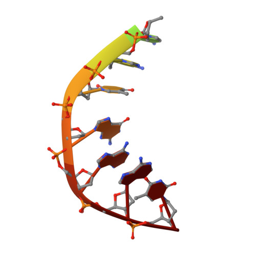
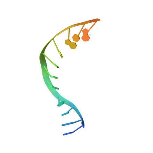
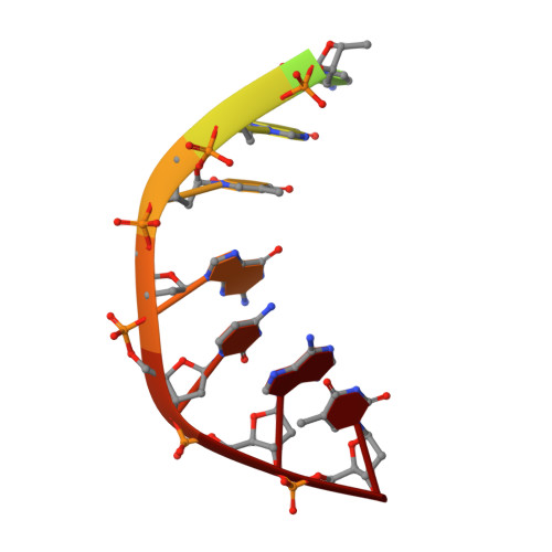
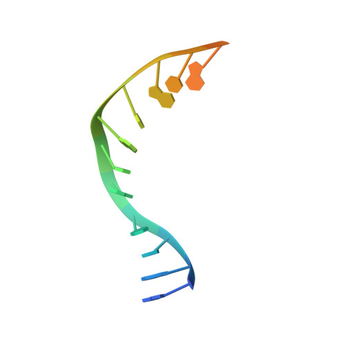
    Type II topoisomerases regulate DNA supercoiling and chromosome segregation. They act as ATP-operated clamps that capture a DNA duplex and pass it through a transient DNA break in a second DNA segment via the sequential opening and closure of ATPase-, G-DNA- and C-gates. Here, we present the first 'open clamp' structures of a 3-gate topoisomerase II-DNA complex, the seminal complex engaged in DNA recognition and capture. A high-resolution structure was solved for a (full-length ParE-ParC55)2 dimer of Streptococcus pneumoniae topoisomerase IV bound to two DNA molecules: a closed DNA gate in a B-A-B form double-helical conformation and a second B-form duplex associated with closed C-gate helices at a novel site neighbouring the catalytically important β-pinwheel DNA-binding domain. The protein N gate is present in an 'arms-wide-open' state with the undimerized N-terminal ParE ATPase domains connected to TOPRIM domains via a flexible joint and folded back allowing ready access both for gate and transported DNA segments and cleavage-stabilizing antibacterial drugs. The structure shows the molecular conformations of all three gates at 3.7 Å, the highest resolution achieved for the full complex to date, and illuminates the mechanism of DNA capture and transport by a type II topoisomerase.


  • Organizational Affiliation

    Randall Division of Cell and Molecular Biophysics, 3rd floor New Hunt's House, Guy's Campus, King's College London, London, SE1 1UL, UK and Division of Biomedical Sciences, St. George's, University of London, Cranmer Terrace, London, SW17 0RE, UK.


Macromolecules

Find similar proteins by:  (by identity cutoff)  |  3D Structure
Entity ID: 1
MoleculeChains Sequence LengthOrganismDetailsImage
DNA topoisomerase 4 subunit A496Streptococcus pneumoniae TIGR4Mutation(s): 0 
Gene Names: parCSP_0855
EC: 5.99.1.3
UniProt
Find proteins for P72525 (Streptococcus pneumoniae serotype 4 (strain ATCC BAA-334 / TIGR4))
Explore P72525 
Go to UniProtKB:  P72525
Entity Groups  
Sequence Clusters30% Identity50% Identity70% Identity90% Identity95% Identity100% Identity
UniProt GroupP72525
Sequence Annotations
Expand
  • Reference Sequence
Find similar proteins by:  (by identity cutoff)  |  3D Structure
Entity ID: 2
MoleculeChains Sequence LengthOrganismDetailsImage
DNA topoisomerase 4 subunit BB [auth C]670Streptococcus pneumoniae TIGR4Mutation(s): 0 
Gene Names: parE
EC: 5.99.1.3
UniProt
Find proteins for Q59961 (Streptococcus pneumoniae serotype 4 (strain ATCC BAA-334 / TIGR4))
Explore Q59961 
Go to UniProtKB:  Q59961
Entity Groups  
Sequence Clusters30% Identity50% Identity70% Identity90% Identity95% Identity100% Identity
UniProt GroupQ59961
Sequence Annotations
Expand
  • Reference Sequence

Find similar nucleic acids by:  Sequence   |   3D Structure  

Entity ID: 3
MoleculeChains LengthOrganismImage
E-site DNAC [auth E]11N/A
Sequence Annotations
Expand
  • Reference Sequence

Find similar nucleic acids by:  Sequence   |   3D Structure  

Entity ID: 4
MoleculeChains LengthOrganismImage
E-site DNAD [auth F]15N/A
Sequence Annotations
Expand
  • Reference Sequence

Find similar nucleic acids by:  Sequence   |   3D Structure  

Entity ID: 5
MoleculeChains LengthOrganismImage
E-site DNAE [auth G]11N/A
Sequence Annotations
Expand
  • Reference Sequence

Find similar nucleic acids by:  Sequence   |   3D Structure  

Entity ID: 6
MoleculeChains LengthOrganismImage
E-site DNAF [auth H]15N/A
Sequence Annotations
Expand
  • Reference Sequence
Small Molecules
Ligands 2 Unique
IDChains Name / Formula / InChI Key2D Diagram3D Interactions
LFX
Query on LFX

Download Ideal Coordinates CCD File 
I [auth F](3S)-9-fluoro-3-methyl-10-(4-methylpiperazin-1-yl)-7-oxo-2,3-dihydro-7H-[1,4]oxazino[2,3,4-ij]quinoline-6-carboxylic acid
C18 H20 F N3 O4
GSDSWSVVBLHKDQ-JTQLQIEISA-N
MG
Query on MG

Download Ideal Coordinates CCD File 
G [auth A],
H [auth C]
MAGNESIUM ION
Mg
JLVVSXFLKOJNIY-UHFFFAOYSA-N
Experimental Data & Validation

Experimental Data

  • Method: X-RAY DIFFRACTION
  • Resolution: 6.53 Å
  • R-Value Free: 0.298 
  • R-Value Work: 0.247 
  • R-Value Observed: 0.250 
  • Space Group: H 3 2
Unit Cell:
Length ( Å )Angle ( ˚ )
a = 213.58α = 90
b = 213.58β = 90
c = 211.83γ = 120
Software Package:
Software NamePurpose
GDEdata collection
PHASERphasing
PHENIXrefinement
xia2data reduction
XDSdata reduction
xia2data scaling
SCALAdata scaling

Structure Validation

View Full Validation Report


Entry History 

Deposition Data

Revision History  (Full details and data files)

  • Version 1.0: 2013-08-28
    Type: Initial release
  • Version 1.1: 2013-09-04
    Changes: Database references
  • Version 1.2: 2013-12-04
    Changes: Database references
  • Version 1.3: 2017-11-15
    Changes: Refinement description
  • Version 1.4: 2023-09-20
    Changes: Data collection, Database references, Derived calculations, Refinement description