4IJJ

Structure of transcription factor DksA2 from Pseudomonas aeruginosa


Experimental Data Snapshot

  • Method: X-RAY DIFFRACTION
  • Resolution: 3.25 Å
  • R-Value Free: 0.310 
  • R-Value Work: 0.257 
  • R-Value Observed: 0.259 

wwPDB Validation   3D Report Full Report


This is version 1.1 of the entry. See complete history


Literature

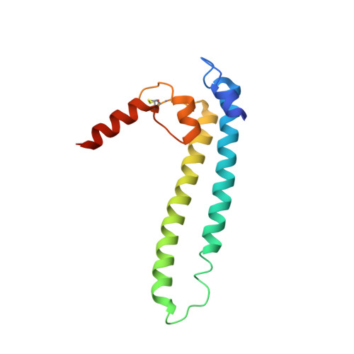
DksA2, a zinc-independent structural analog of the transcription factor DksA.

Furman, R.Biswas, T.Danhart, E.M.Foster, M.P.Tsodikov, O.V.Artsimovitch, I.

(2013) FEBS Lett 587: 614-619

  • DOI: https://doi.org/10.1016/j.febslet.2013.01.073
  • Primary Citation of Related Structures:  
    4IJJ

  • PubMed Abstract: 

    Transcription factor DksA contains a four-Cys Zn(2 +)-finger motif thought to be responsible for structural integrity and the relative disposition of its domains. Pseudomonas aeruginosa encodes an additional DksA paralog (DksA2) that is expressed selectively under Zn(2+) limitation. Although DksA2 does not bind Zn(2+), it complements the Escherichia coli dksA deletion and has similar effects on transcription in vitro. In this study, structural and biochemical analyses reveal that DksA2 has a similar fold, domain structure and RNA polymerase binding properties to those of the E. coli DksA despite the lack of the stabilizing metal ion.


  • Organizational Affiliation

    Department of Microbiology, The Ohio State University, Columbus, OH, USA.


Macromolecules
Find similar proteins by:  (by identity cutoff)  |  3D Structure
Entity ID: 1
MoleculeChains Sequence LengthOrganismDetailsImage
Putative C4-type zinc finger protein, DksA/TraR family
A, B, C
136Pseudomonas aeruginosa UCBPP-PA14Mutation(s): 0 
Gene Names: PA14_73020
UniProt
Find proteins for A0A0H2ZJP8 (Pseudomonas aeruginosa (strain UCBPP-PA14))
Explore A0A0H2ZJP8 
Go to UniProtKB:  A0A0H2ZJP8
Entity Groups  
Sequence Clusters30% Identity50% Identity70% Identity90% Identity95% Identity100% Identity
UniProt GroupA0A0H2ZJP8
Sequence Annotations
Expand
  • Reference Sequence
Experimental Data & Validation

Experimental Data

  • Method: X-RAY DIFFRACTION
  • Resolution: 3.25 Å
  • R-Value Free: 0.310 
  • R-Value Work: 0.257 
  • R-Value Observed: 0.259 
  • Space Group: P 6 2 2
Unit Cell:
Length ( Å )Angle ( ˚ )
a = 169.453α = 90
b = 169.453β = 90
c = 92.32γ = 120
Software Package:
Software NamePurpose
HKL-2000data collection
PHASERphasing
REFMACrefinement
HKL-2000data reduction
HKL-2000data scaling

Structure Validation

View Full Validation Report


Entry History 

Deposition Data

Revision History  (Full details and data files)

  • Version 1.0: 2013-02-27
    Type: Initial release
  • Version 1.1: 2013-03-27
    Changes: Database references