4I53

Crystal structure of clade C1086 HIV-1 gp120 core in complex with DMJ-II-121


Experimental Data Snapshot

  • Method: X-RAY DIFFRACTION
  • Resolution: 2.50 Å
  • R-Value Free: 0.273 
  • R-Value Work: 0.254 
  • R-Value Observed: 0.255 

wwPDB Validation   3D Report Full Report


Ligand Structure Quality Assessment 


This is version 1.2 of the entry. See complete history


Literature

Structure-Based Design and Synthesis of an HIV-1 Entry Inhibitor Exploiting X-Ray and Thermodynamic Characterization.

Lalonde, J.M.Le-Khac, M.Jones, D.M.Courter, J.R.Park, J.Schon, A.Princiotto, A.M.Wu, X.Mascola, J.R.Freire, E.Sodroski, J.Madani, N.Hendrickson, W.A.Smith, A.B.

(2013) ACS Med Chem Lett 4: 338-343

  • DOI: https://doi.org/10.1021/ml300407y
  • Primary Citation of Related Structures:  
    4I53, 4I54

  • PubMed Abstract: 

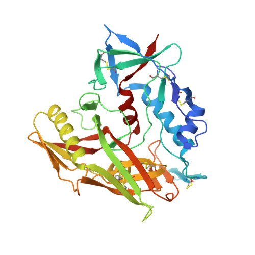
    The design, synthesis, thermodynamic and crystallographic characterization of a potent, broad spectrum, second-generation HIV-1 entry inhibitor that engages conserved carbonyl hydrogen bonds within gp120 has been achieved. The optimized antagonist exhibits a sub-micromolar binding affinity (110 nM) and inhibits viral entry of clade B and C viruses (IC 50 geometric mean titer of 1.7 and 14.0 μM, respectively), without promoting CD4-independent viral entry. thermodynamic signatures indicate a binding preference for the ( R,R )-over the ( S,S )-enantiomer. The crystal structure of the small molecule-gp120 complex reveals the displacement of crystallographic water and the formation of a hydrogen bond with a backbone carbonyl of the bridging sheet. Thus, structure-based design and synthesis targeting the highly conserved and structurally characterized CD4:gp120 interface is an effective tactic to enhance the neutralization potency of small molecule HIV-1 entry inhibitors.


  • Organizational Affiliation

    Department of Chemistry, Bryn Mawr College, Bryn Mawr, PA 19010.


Macromolecules
Find similar proteins by:  (by identity cutoff)  |  3D Structure
Entity ID: 1
MoleculeChains Sequence LengthOrganismDetailsImage
HIV-1 glycoprotein
A, B
358Human immunodeficiency virus 1Mutation(s): 0 
UniProt
Find proteins for C6G099 (Human immunodeficiency virus 1)
Explore C6G099 
Go to UniProtKB:  C6G099
Entity Groups  
Sequence Clusters30% Identity50% Identity70% Identity90% Identity95% Identity100% Identity
UniProt GroupC6G099
Sequence Annotations
Expand
  • Reference Sequence
Small Molecules
Ligands 3 Unique
IDChains Name / Formula / InChI Key2D Diagram3D Interactions
1C1
Query on 1C1

Download Ideal Coordinates CCD File 
C [auth A],
K [auth B]
amino({[(1R,2R)-1-({[(4-chloro-3-fluorophenyl)amino](oxo)acetyl}amino)-2,3-dihydro-1H-inden-2-yl]methyl}amino)methanimi nium
C19 H20 Cl F N5 O2
PZYMVIYVOCOSHZ-BDJLRTHQSA-O
NAG
Query on NAG

Download Ideal Coordinates CCD File 
D [auth A]
E [auth A]
F [auth A]
G [auth A]
H [auth A]
D [auth A],
E [auth A],
F [auth A],
G [auth A],
H [auth A],
I [auth A],
J [auth A],
M [auth B],
N [auth B],
O [auth B],
P [auth B],
Q [auth B],
R [auth B]
2-acetamido-2-deoxy-beta-D-glucopyranose
C8 H15 N O6
OVRNDRQMDRJTHS-FMDGEEDCSA-N
FMT
Query on FMT

Download Ideal Coordinates CCD File 
L [auth B]FORMIC ACID
C H2 O2
BDAGIHXWWSANSR-UHFFFAOYSA-N
Binding Affinity Annotations 
IDSourceBinding Affinity
1C1 Binding MOAD:  4I53 Kd: 110 (nM) from 1 assay(s)
Experimental Data & Validation

Experimental Data

  • Method: X-RAY DIFFRACTION
  • Resolution: 2.50 Å
  • R-Value Free: 0.273 
  • R-Value Work: 0.254 
  • R-Value Observed: 0.255 
  • Space Group: C 2 2 21
Unit Cell:
Length ( Å )Angle ( ˚ )
a = 67.45α = 90
b = 127.67β = 90
c = 192.9γ = 90
Software Package:
Software NamePurpose
PHENIXrefinement

Structure Validation

View Full Validation Report


Ligand Structure Quality Assessment 


Entry History 

Deposition Data

Revision History  (Full details and data files)

  • Version 1.0: 2013-05-29
    Type: Initial release
  • Version 1.1: 2020-07-29
    Type: Remediation
    Reason: Carbohydrate remediation
    Changes: Data collection, Derived calculations, Structure summary
  • Version 1.2: 2021-05-26
    Changes: Source and taxonomy, Structure summary