4HE4

Crystal structure of the yellow fluorescent protein phiYFP (Phialidium sp.)


Experimental Data Snapshot

  • Method: X-RAY DIFFRACTION
  • Resolution: 2.05 Å
  • R-Value Free: 0.256 
  • R-Value Work: 0.196 
  • R-Value Observed: 0.199 

wwPDB Validation   3D Report Full Report


This is version 1.3 of the entry. See complete history


Literature

Yellow fluorescent protein phiYFPv (Phialidium): structure and structure-based mutagenesis.

Pletneva, N.V.Pletnev, V.Z.Souslova, E.Chudakov, D.M.Lukyanov, S.Martynov, V.I.Arhipova, S.Artemyev, I.Wlodawer, A.Dauter, Z.Pletnev, S.

(2013) Acta Crystallogr D Biol Crystallogr 69: 1005-1012

  • DOI: https://doi.org/10.1107/S0907444913004034
  • Primary Citation of Related Structures:  
    4HE4

  • PubMed Abstract: 

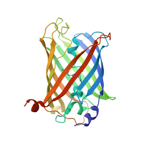
    The yellow fluorescent protein phiYFPv (λem(max) ≃ 537 nm) with improved folding has been developed from the spectrally identical wild-type phiYFP found in the marine jellyfish Phialidium. The latter fluorescent protein is one of only two known cases of naturally occurring proteins that exhibit emission spectra in the yellow-orange range (535-555 nm). Here, the crystal structure of phiYFPv has been determined at 2.05 Å resolution. The `yellow' chromophore formed from the sequence triad Thr65-Tyr66-Gly67 adopts the bicyclic structure typical of fluorophores emitting in the green spectral range. It was demonstrated that perfect antiparallel π-stacking of chromophore Tyr66 and the proximal Tyr203, as well as Val205, facing the chromophore phenolic ring are chiefly responsible for the observed yellow emission of phiYFPv at 537 nm. Structure-based site-directed mutagenesis has been used to identify the key functional residues in the chromophore environment. The obtained results have been utilized to improve the properties of phiYFPv and its homologous monomeric biomarker tagYFP.


  • Organizational Affiliation

    Shemyakin-Ovchinnikov Institute of Bioorganic Chemistry, Russian Academy of Sciences, Moscow, Russian Federation.


Macromolecules
Find similar proteins by:  (by identity cutoff)  |  3D Structure
Entity ID: 1
MoleculeChains Sequence LengthOrganismDetailsImage
Yellow fluorescent protein
A, B
244Phialidium sp. SL-2003Mutation(s): 1 
UniProt
Find proteins for Q6RYS7 (Phialidium sp. SL-2003)
Explore Q6RYS7 
Go to UniProtKB:  Q6RYS7
Entity Groups  
Sequence Clusters30% Identity50% Identity70% Identity90% Identity95% Identity100% Identity
UniProt GroupQ6RYS7
Sequence Annotations
Expand
  • Reference Sequence
Small Molecules
Modified Residues  1 Unique
IDChains TypeFormula2D DiagramParent
CRO
Query on CRO
A, B
L-PEPTIDE LINKINGC15 H17 N3 O5THR, TYR, GLY
Experimental Data & Validation

Experimental Data

  • Method: X-RAY DIFFRACTION
  • Resolution: 2.05 Å
  • R-Value Free: 0.256 
  • R-Value Work: 0.196 
  • R-Value Observed: 0.199 
  • Space Group: H 3 2
Unit Cell:
Length ( Å )Angle ( ˚ )
a = 102.87α = 90
b = 102.87β = 90
c = 242.53γ = 120
Software Package:
Software NamePurpose
SCALEPACKdata scaling
REFMACrefinement
PDB_EXTRACTdata extraction
SERGUIdata collection
HKL-2000data reduction
HKL-2000data scaling
MOLREPphasing

Structure Validation

View Full Validation Report


Entry History 

Deposition Data

Revision History  (Full details and data files)

  • Version 1.0: 2013-05-29
    Type: Initial release
  • Version 1.1: 2013-06-19
    Changes: Database references
  • Version 1.2: 2023-09-20
    Changes: Data collection, Database references, Derived calculations, Refinement description
  • Version 1.3: 2023-12-06
    Changes: Data collection