4H8K

Crystal structure of LC11-RNase H1 in complex with RNA/DNA hybrid


Experimental Data Snapshot

  • Method: X-RAY DIFFRACTION
  • Resolution: 2.30 Å
  • R-Value Free: 0.278 
  • R-Value Work: 0.207 
  • R-Value Observed: 0.210 

wwPDB Validation   3D Report Full Report


This is version 1.1 of the entry. See complete history


Literature

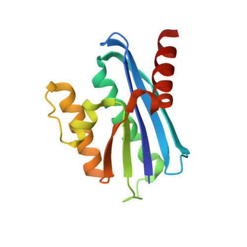
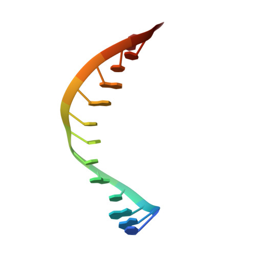
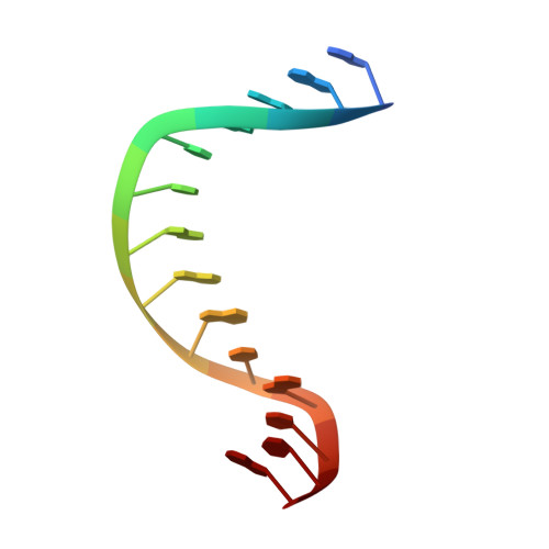
Crystal structure of metagenome-derived LC11-RNase H1 in complex with RNA/DNA hybrid

Nguyen, T.N.You, D.J.Matsumoto, H.Kanaya, E.Koga, Y.Kanaya, S.

(2013) J Struct Biol 182: 144-154

  • DOI: https://doi.org/10.1016/j.jsb.2013.02.018
  • Primary Citation of Related Structures:  
    4H8K

  • PubMed Abstract: 

    LC11-RNase H1 is a Sulfolobus tokodaii RNase H1 (Sto-RNase H1) homologue isolated by metagenomic approach. In this study, the crystal structure of LC11-RNase H1 in complex with an RNA/DNA substrate was determined. Unlike Bacillus halodurans RNase H1 without hybrid binding domain (HBD) (Bh-RNase HC) and human RNase H1 without HBD (Hs-RNase HC), LC11-RNase H1 interacts with four non-consecutive 2'-OH groups of the RNA strand. The lack of interactions with four consecutive 2'-OH groups leads to a dramatic decrease in the ability of LC11-RNase H1 to cleave the DNA-RNA-DNA/DNA substrate containing four ribonucleotides as compared to those to cleave the substrates containing five and six ribonucleotides. The interaction of LC11-RNase H1 with the DNA strand is also different from those of Bh-RNase HC and Hs-RNase HC. Beside the common phosphate-binding pocket, LC11-RNase H1 has a unique DNA-binding channel. Furthermore, the active-site residues of LC11-RNase H1 are located farther away from the scissile phosphate group than those of Bh-RNase HC and Hs-RNase HC. Modeling of Sto-RNase H1 in complex with the 14bp RNA/DNA substrate, together with the structure-based mutational analyses, suggest that the ability of Sto-RNase H1 to cleave double-stranded RNA is dependent on the local conformation of the basic residues located at the DNA binding site.


  • Organizational Affiliation

    Department of Material and Life Science, Graduate School of Engineering, Osaka University, 2-1 Yamadaoka, Suita, Osaka 565-0871, Japan.


Macromolecules

Find similar proteins by:  (by identity cutoff)  |  3D Structure
Entity ID: 1
MoleculeChains Sequence LengthOrganismDetailsImage
Ribonuclease H
A, B
140uncultured organismMutation(s): 1 
EC: 3.1.26.4
UniProt
Find proteins for E0X767 (uncultured organism)
Explore E0X767 
Go to UniProtKB:  E0X767
Entity Groups  
Sequence Clusters30% Identity50% Identity70% Identity90% Identity95% Identity100% Identity
UniProt GroupE0X767
Sequence Annotations
Expand
  • Reference Sequence

Find similar nucleic acids by:  Sequence   |   3D Structure  

Entity ID: 2
MoleculeChains LengthOrganismImage
RNA (5'-R(*CP*GP*AP*CP*AP*CP*CP*UP*GP*AP*UP*UP*CP*C)-3')14N/A
Sequence Annotations
Expand
  • Reference Sequence

Find similar nucleic acids by:  Sequence   |   3D Structure  

Entity ID: 3
MoleculeChains LengthOrganismImage
DNA (5'-D(*GP*GP*AP*AP*TP*CP*AP*GP*GP*TP*GP*TP*CP*G)-3')14N/A
Sequence Annotations
Expand
  • Reference Sequence
Experimental Data & Validation

Experimental Data

  • Method: X-RAY DIFFRACTION
  • Resolution: 2.30 Å
  • R-Value Free: 0.278 
  • R-Value Work: 0.207 
  • R-Value Observed: 0.210 
  • Space Group: P 1 21 1
Unit Cell:
Length ( Å )Angle ( ˚ )
a = 42.613α = 90
b = 90.758β = 97.82
c = 55.605γ = 90
Software Package:
Software NamePurpose
HKL-2000data collection
MOLREPphasing
REFMACrefinement
HKL-2000data reduction
HKL-2000data scaling

Structure Validation

View Full Validation Report


Entry History 

Deposition Data

Revision History  (Full details and data files)

  • Version 1.0: 2013-09-25
    Type: Initial release
  • Version 1.1: 2023-11-08
    Changes: Data collection, Database references, Refinement description