Human UDP-alpha-d-xylose Synthase and Escherichia coli ArnA Conserve a Conformational Shunt That Controls Whether Xylose or 4-Keto-Xylose Is Produced.
Polizzi, S.J., Walsh Jr., R.M., Peeples, W.B., Lim, J.M., Wells, L., Wood, Z.A.(2012) Biochemistry 51: 8844-8855
- PubMed: 23072385
- DOI: https://doi.org/10.1021/bi301135b
- Primary Citation of Related Structures:
4GLL - PubMed Abstract:
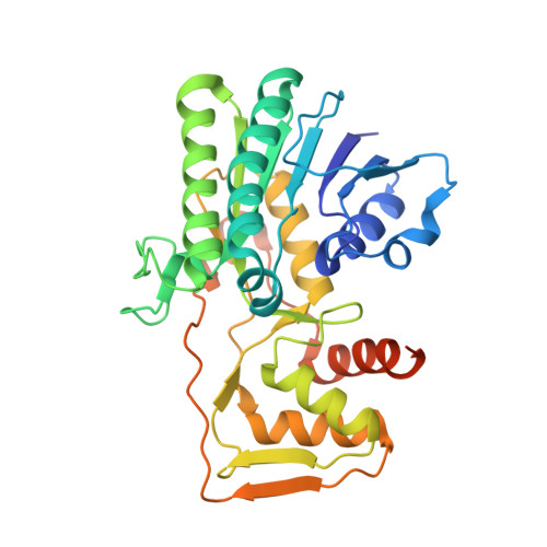
Human UDP-α-D-xylose synthase (hUXS) is a member of the short-chain dehydrogenase/reductase family of nucleotide-sugar modifying enzymes. hUXS contains a bound NAD(+) cofactor that it recycles by first oxidizing UDP-α-D-glucuronic acid (UGA), and then reducing the UDP-α-D-4-keto-xylose (UX4O) to produce UDP-α-D-xylose (UDX). Despite the observation that purified hUXS contains a bound cofactor, it has been reported that exogenous NAD(+) will stimulate enzyme activity. Here we show that a small fraction of hUXS releases the NADH and UX4O intermediates as products during turnover. The resulting apoenzyme can be rescued by exogenous NAD(+), explaining the apparent stimulatory effect of added cofactor. The slow release of NADH and UX4O as side products by hUXS is reminiscent of the Escherichia coli UGA decarboxylase (ArnA), a related enzyme that produces NADH and UX4O as products. We report that ArnA can rebind NADH and UX4O to slowly make UDX. This means that both enzymes share the same catalytic machinery, but differ in the preferred final product. We present a bifurcated rate equation that explains how the substrate is shunted to the distinct final products. Using a new crystal structure of hUXS, we identify the structural elements of the shunt and propose that the local unfolding of the active site directs reactants toward the preferred products. Finally, we present evidence that the release of NADH and UX4O involves a cooperative conformational change that is conserved in both enzymes.
Organizational Affiliation:
Department of Biochemistry and Molecular Biology, University of Georgia, Athens, Georgia 30602, United States.