4DS7

Crystal structure of yeast calmodulin bound to the C-terminal fragment of spindle pole body protein Spc110


Experimental Data Snapshot

  • Method: X-RAY DIFFRACTION
  • Resolution: 2.15 Å
  • R-Value Free: 0.240 
  • R-Value Work: 0.197 
  • R-Value Observed: 0.199 

wwPDB Validation   3D Report Full Report


This is version 1.3 of the entry. See complete history


Literature

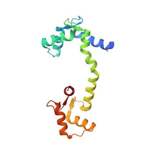
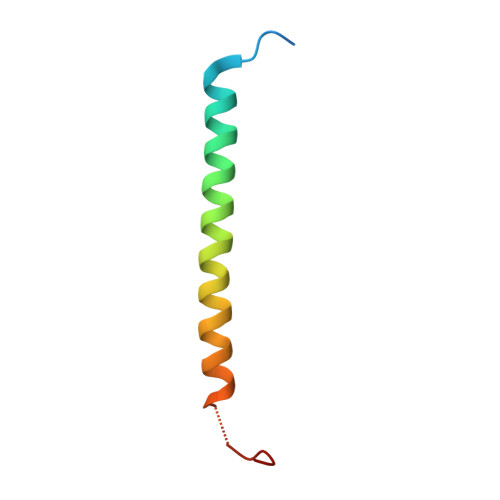
The molecular architecture of the yeast spindle pole body core determined by Bayesian integrative modeling.

Viswanath, S.Bonomi, M.Kim, S.J.Klenchin, V.A.Taylor, K.C.Yabut, K.C.Umbreit, N.T.Van Epps, H.A.Meehl, J.Jones, M.H.Russel, D.Velazquez-Muriel, J.A.Winey, M.Rayment, I.Davis, T.N.Sali, A.Muller, E.G.

(2017) Mol Biol Cell 28: 3298-3314

  • DOI: https://doi.org/10.1091/mbc.E17-06-0397
  • Primary Citation of Related Structures:  
    4DS7

  • PubMed Abstract: 

    Microtubule-organizing centers (MTOCs) form, anchor, and stabilize the polarized network of microtubules in a cell. The central MTOC is the centrosome that duplicates during the cell cycle and assembles a bipolar spindle during mitosis to capture and segregate sister chromatids. Yet, despite their importance in cell biology, the physical structure of MTOCs is poorly understood. Here we determine the molecular architecture of the core of the yeast spindle pole body (SPB) by Bayesian integrative structure modeling based on in vivo fluorescence resonance energy transfer (FRET), small-angle x-ray scattering (SAXS), x-ray crystallography, electron microscopy, and two-hybrid analysis. The model is validated by several methods that include a genetic analysis of the conserved PACT domain that recruits Spc110, a protein related to pericentrin, to the SPB. The model suggests that calmodulin can act as a protein cross-linker and Spc29 is an extended, flexible protein. The model led to the identification of a single, essential heptad in the coiled-coil of Spc110 and a minimal PACT domain. It also led to a proposed pathway for the integration of Spc110 into the SPB.


  • Organizational Affiliation

    Department of Bioengineering and Therapeutic Sciences, Department of Pharmaceutical Chemistry, California Institute for Quantitative Biosciences, University of California, San Francisco, San Francisco, CA 94158.


Macromolecules
Find similar proteins by:  (by identity cutoff)  |  3D Structure
Entity ID: 1
MoleculeChains Sequence LengthOrganismDetailsImage
Calmodulin
A, B, C, D
147Kluyveromyces lactis NRRL Y-1140Mutation(s): 0 
Gene Names: CMD1KLLA0B09427g
UniProt
Find proteins for O60041 (Kluyveromyces lactis (strain ATCC 8585 / CBS 2359 / DSM 70799 / NBRC 1267 / NRRL Y-1140 / WM37))
Explore O60041 
Go to UniProtKB:  O60041
Entity Groups  
Sequence Clusters30% Identity50% Identity70% Identity90% Identity95% Identity100% Identity
UniProt GroupO60041
Sequence Annotations
Expand
  • Reference Sequence
Find similar proteins by:  (by identity cutoff)  |  3D Structure
Entity ID: 2
MoleculeChains Sequence LengthOrganismDetailsImage
Spindle pole body component 110
E, F, G, H
55Saccharomyces cerevisiae S288CMutation(s): 0 
Gene Names: SPC110NUF1XCM1YDR356WD9476.3
UniProt
Find proteins for P32380 (Saccharomyces cerevisiae (strain ATCC 204508 / S288c))
Explore P32380 
Go to UniProtKB:  P32380
Entity Groups  
Sequence Clusters30% Identity50% Identity70% Identity90% Identity95% Identity100% Identity
UniProt GroupP32380
Sequence Annotations
Expand
  • Reference Sequence
Small Molecules
Ligands 3 Unique
IDChains Name / Formula / InChI Key2D Diagram3D Interactions
SO4
Query on SO4

Download Ideal Coordinates CCD File 
L [auth A]SULFATE ION
O4 S
QAOWNCQODCNURD-UHFFFAOYSA-L
GOL
Query on GOL

Download Ideal Coordinates CCD File 
K [auth A],
O [auth B],
P [auth B],
Q [auth B]
GLYCEROL
C3 H8 O3
PEDCQBHIVMGVHV-UHFFFAOYSA-N
SR
Query on SR

Download Ideal Coordinates CCD File 
I [auth A]
J [auth A]
M [auth B]
N [auth B]
R [auth C]
I [auth A],
J [auth A],
M [auth B],
N [auth B],
R [auth C],
S [auth C],
T [auth D],
U [auth D]
STRONTIUM ION
Sr
PWYYWQHXAPXYMF-UHFFFAOYSA-N
Experimental Data & Validation

Experimental Data

  • Method: X-RAY DIFFRACTION
  • Resolution: 2.15 Å
  • R-Value Free: 0.240 
  • R-Value Work: 0.197 
  • R-Value Observed: 0.199 
  • Space Group: P 21 21 2
Unit Cell:
Length ( Å )Angle ( ˚ )
a = 114.67α = 90
b = 145.444β = 90
c = 49.837γ = 90
Software Package:
Software NamePurpose
DENZOdata reduction
SCALEPACKdata scaling
MOLREPphasing
REFMACrefinement
PDB_EXTRACTdata extraction
d*TREKdata scaling
HKL-3000data reduction
HKL-3000data scaling

Structure Validation

View Full Validation Report


Entry History 

Deposition Data

Revision History  (Full details and data files)

  • Version 1.0: 2012-03-07
    Type: Initial release
  • Version 1.1: 2017-10-04
    Changes: Database references, Refinement description
  • Version 1.2: 2023-07-26
    Changes: Database references, Derived calculations
  • Version 1.3: 2023-09-13
    Changes: Data collection, Refinement description