4D1Q

Hermes transposase bound to its terminal inverted repeat


Experimental Data Snapshot

  • Method: X-RAY DIFFRACTION
  • Resolution: 3.40 Å
  • R-Value Free: 0.254 
  • R-Value Work: 0.212 
  • R-Value Observed: 0.212 

wwPDB Validation   3D Report Full Report


This is version 1.1 of the entry. See complete history


Literature

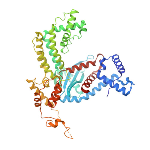
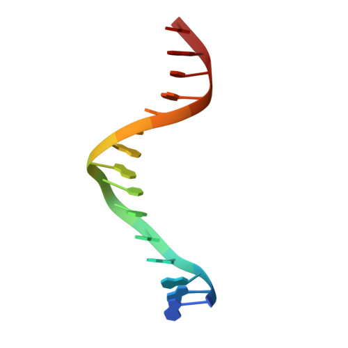
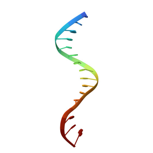
Structural Basis of Hat Transposon End Recognition by Hermes, an Octameric DNA Transposase from Musca Domestica.

Hickman, A.B.Ewis, H.E.Li, X.Knapp, J.A.Laver, T.Doss, A.Tolun, G.Steven, A.C.Grishaev, A.Bax, A.Atkinson, P.W.Craig, N.L.Dyda, F.

(2014) Cell 158: 353

  • DOI: https://doi.org/10.1016/j.cell.2014.05.037
  • Primary Citation of Related Structures:  
    4D1Q

  • PubMed Abstract: 

    Hermes is a member of the hAT transposon superfamily that has active representatives, including McClintock's archetypal Ac mobile genetic element, in many eukaryotic species. The crystal structure of the Hermes transposase-DNA complex reveals that Hermes forms an octameric ring organized as a tetramer of dimers. Although isolated dimers are active in vitro for all the chemical steps of transposition, only octamers are active in vivo. The octamer can provide not only multiple specific DNA-binding domains to recognize repeated subterminal sequences within the transposon ends, which are important for activity, but also multiple nonspecific DNA binding surfaces for target capture. The unusual assembly explains the basis of bipartite DNA recognition at hAT transposon ends, provides a rationale for transposon end asymmetry, and suggests how the avidity provided by multiple sites of interaction could allow a transposase to locate its transposon ends amidst a sea of chromosomal DNA.


  • Organizational Affiliation

    Laboratory of Molecular Biology, National Institute of Diabetes and Digestive and Kidney Diseases, National Institutes of Health, Bethesda, MD 20892, USA.


Macromolecules

Find similar proteins by:  (by identity cutoff)  |  3D Structure
Entity ID: 1
MoleculeChains Sequence LengthOrganismDetailsImage
TRANSPOSASE
A, B, G, H
536Musca domesticaMutation(s): 2 
UniProt
Find proteins for Q25442 (Musca domestica)
Explore Q25442 
Go to UniProtKB:  Q25442
Entity Groups  
Sequence Clusters30% Identity50% Identity70% Identity90% Identity95% Identity100% Identity
UniProt GroupQ25442
Sequence Annotations
Expand
  • Reference Sequence

Find similar nucleic acids by:  Sequence   |   3D Structure  

Entity ID: 2
MoleculeChains LengthOrganismImage
TERMINAL INVERTED REPEAT
C, E, I, K
15Musca domestica
Sequence Annotations
Expand
  • Reference Sequence

Find similar nucleic acids by:  Sequence   |   3D Structure  

Entity ID: 3
MoleculeChains LengthOrganismImage
TERMINAL INVERTED REPEAT
D, F, J, L
16Musca domestica
Sequence Annotations
Expand
  • Reference Sequence
Experimental Data & Validation

Experimental Data

  • Method: X-RAY DIFFRACTION
  • Resolution: 3.40 Å
  • R-Value Free: 0.254 
  • R-Value Work: 0.212 
  • R-Value Observed: 0.212 
  • Space Group: P 32 2 1
Unit Cell:
Length ( Å )Angle ( ˚ )
a = 265.15α = 90
b = 265.15β = 90
c = 157.64γ = 120
Software Package:
Software NamePurpose
XDSdata reduction
XDSdata scaling
SHARPphasing
DMphasing
CNSrefinement

Structure Validation

View Full Validation Report


Entry History 

Deposition Data

Revision History  (Full details and data files)

  • Version 1.0: 2014-07-30
    Type: Initial release
  • Version 1.1: 2017-06-28
    Changes: Data collection