4BOH

Madanins (MEROPS I53) are cleaved by thrombin and factor Xa


Experimental Data Snapshot

  • Method: X-RAY DIFFRACTION
  • Resolution: 2.60 Å
  • R-Value Free: 0.239 
  • R-Value Work: 0.191 
  • R-Value Observed: 0.193 

wwPDB Validation   3D Report Full Report


Ligand Structure Quality Assessment 


This is version 1.2 of the entry. See complete history


Literature

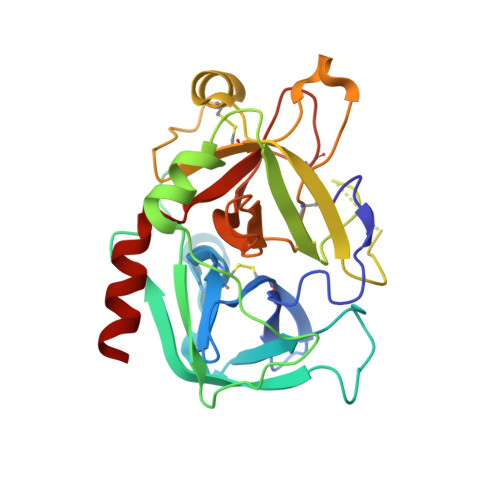
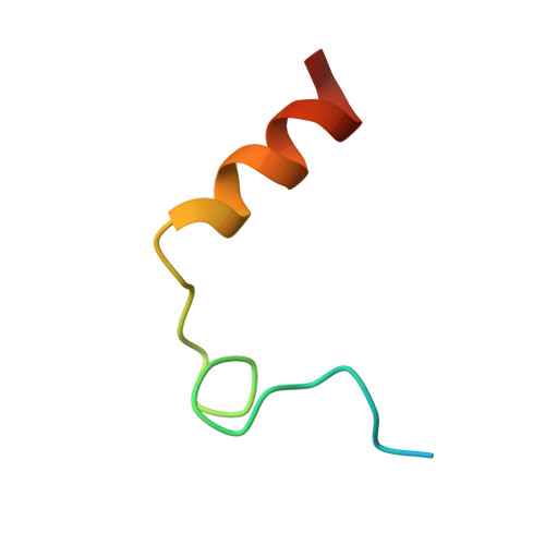
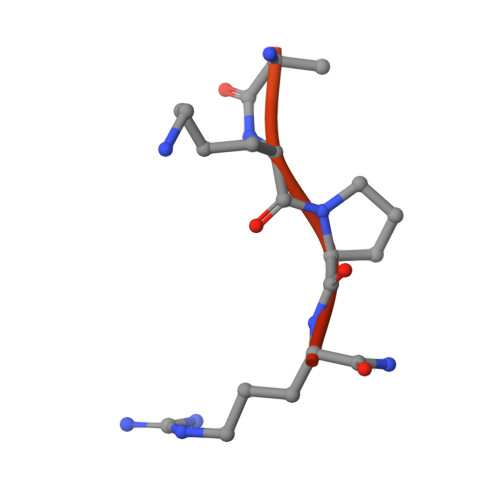
The Tick-Derived Anticoagulant Madanin is Processed by Thrombin and Factor Xa.

Figueiredo, A.C.De Sanctis, D.Pereira, P.J.

(2013) PLoS One 8: 71866

  • DOI: https://doi.org/10.1371/journal.pone.0071866
  • Primary Citation of Related Structures:  
    4BOH

  • PubMed Abstract: 

    The cysteine-less peptidic anticoagulants madanin-1 and madanin-2 from the bush tick Haemaphysalis longicornis are the founding members of the MEROPS inhibitor family I53. It has been previously suggested that madanins exert their functional activity by competing with physiological substrates for binding to the positively charged exosite I (fibrinogen-binding exosite) of α-thrombin. We hereby demonstrate that competitive inhibition of α-thrombin by madanin-1 or madanin-2 involves binding to the enzyme's active site. Moreover, the blood coagulation factors IIa and Xa are shown to hydrolyze both inhibitors at different, although partially overlapping cleavage sites. Finally, the three-dimensional structure of the complex formed between human α-thrombin and a proteolytic fragment of madanin-1, determined by X-ray crystallography, elucidates the molecular details of madanin-1 recognition and processing by the proteinase. Taken together, the current findings establish the mechanism of action of madanins, natural anticoagulants that behave as cleavable competitive inhibitors of thrombin.


  • Organizational Affiliation

    IBMC - Instituto de Biologia Molecular e Celular, Universidade do Porto, Porto, Portugal.


Macromolecules
Find similar proteins by:  (by identity cutoff)  |  3D Structure
Entity ID: 1
MoleculeChains Sequence LengthOrganismDetailsImage
THROMBIN HEAVY CHAINA,
C [auth H]
259Homo sapiensMutation(s): 0 
EC: 3.4.21.5
UniProt & NIH Common Fund Data Resources
Find proteins for P00734 (Homo sapiens)
Explore P00734 
Go to UniProtKB:  P00734
PHAROS:  P00734
GTEx:  ENSG00000180210 
Entity Groups  
Sequence Clusters30% Identity50% Identity70% Identity90% Identity95% Identity100% Identity
UniProt GroupP00734
Sequence Annotations
Expand
  • Reference Sequence
Find similar proteins by:  (by identity cutoff)  |  3D Structure
Entity ID: 2
MoleculeChains Sequence LengthOrganismDetailsImage
THROMBIN LIGHT CHAINB,
D [auth L]
36Homo sapiensMutation(s): 0 
EC: 3.4.21.5
UniProt & NIH Common Fund Data Resources
Find proteins for P00734 (Homo sapiens)
Explore P00734 
Go to UniProtKB:  P00734
PHAROS:  P00734
GTEx:  ENSG00000180210 
Entity Groups  
Sequence Clusters30% Identity50% Identity70% Identity90% Identity95% Identity100% Identity
UniProt GroupP00734
Sequence Annotations
Expand
  • Reference Sequence
Find similar proteins by:  (by identity cutoff)  |  3D Structure
Entity ID: 3
MoleculeChains Sequence LengthOrganismDetailsImage
THROMBIN INHIBITOR MADANIN 1E [auth M]60Haemaphysalis longicornisMutation(s): 0 
UniProt
Find proteins for Q86FP9 (Haemaphysalis longicornis)
Explore Q86FP9 
Go to UniProtKB:  Q86FP9
Entity Groups  
Sequence Clusters30% Identity50% Identity70% Identity90% Identity95% Identity100% Identity
UniProt GroupQ86FP9
Sequence Annotations
Expand
  • Reference Sequence
Small Molecules
Ligands 3 Unique
IDChains Name / Formula / InChI Key2D Diagram3D Interactions
NAG
Query on NAG

Download Ideal Coordinates CCD File 
F [auth A],
K [auth H]
2-acetamido-2-deoxy-beta-D-glucopyranose
C8 H15 N O6
OVRNDRQMDRJTHS-FMDGEEDCSA-N
SO4
Query on SO4

Download Ideal Coordinates CCD File 
H [auth A],
I [auth A],
J [auth A],
M [auth H],
N [auth H]
SULFATE ION
O4 S
QAOWNCQODCNURD-UHFFFAOYSA-L
NA
Query on NA

Download Ideal Coordinates CCD File 
G [auth A],
L [auth H]
SODIUM ION
Na
FKNQFGJONOIPTF-UHFFFAOYSA-N
Experimental Data & Validation

Experimental Data

  • Method: X-RAY DIFFRACTION
  • Resolution: 2.60 Å
  • R-Value Free: 0.239 
  • R-Value Work: 0.191 
  • R-Value Observed: 0.193 
  • Space Group: P 21 21 21
Unit Cell:
Length ( Å )Angle ( ˚ )
a = 53.071α = 90
b = 78.578β = 90
c = 155.156γ = 90
Software Package:
Software NamePurpose
PHENIXrefinement
XDSdata reduction
XSCALEdata scaling
PHASERphasing

Structure Validation

View Full Validation Report


Ligand Structure Quality Assessment 


Entry History 

Deposition Data

Revision History  (Full details and data files)

  • Version 1.0: 2013-09-04
    Type: Initial release
  • Version 1.1: 2020-07-29
    Type: Remediation
    Reason: Carbohydrate remediation
    Changes: Data collection, Derived calculations, Other, Structure summary
  • Version 1.2: 2023-12-20
    Changes: Data collection, Database references, Refinement description, Structure summary