4N8M

Structural polymorphism in the N-terminal oligomerization domain of NPM1


Experimental Data Snapshot

  • Method: X-RAY DIFFRACTION
  • Resolution: 1.80 Å
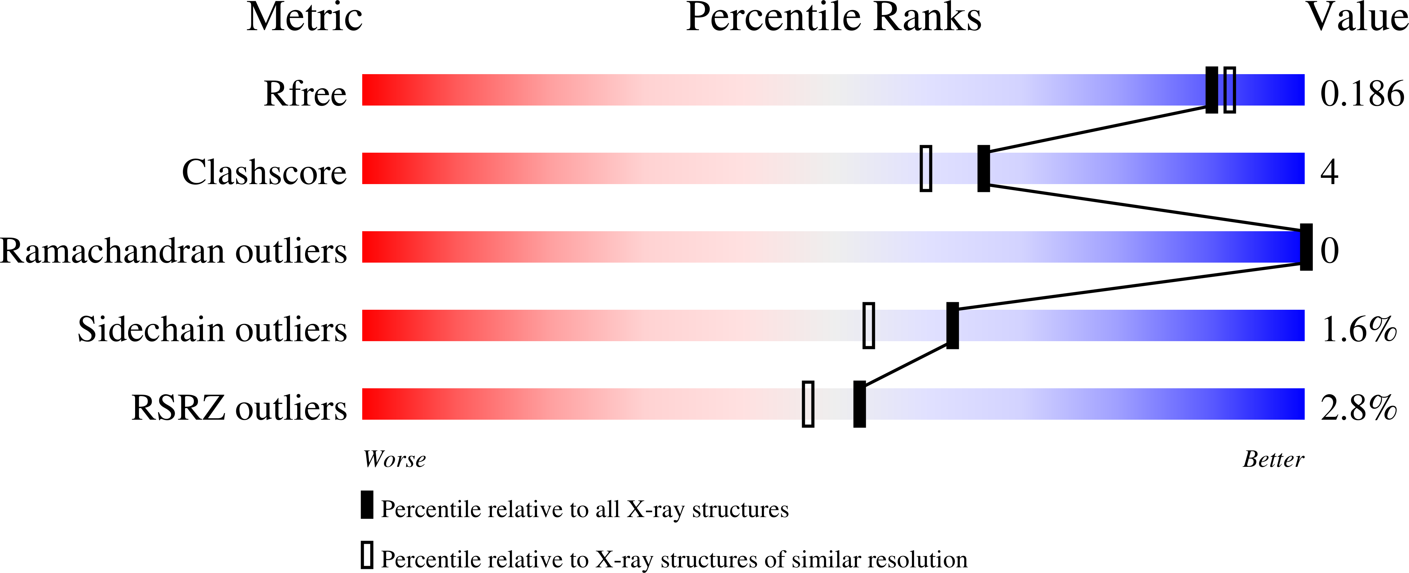
  • R-Value Free: 0.190 
  • R-Value Work: 0.163 
  • R-Value Observed: 0.165 

wwPDB Validation   3D Report Full Report


This is version 1.3 of the entry. See complete history


Literature

Structural polymorphism in the N-terminal oligomerization domain of NPM1.

Mitrea, D.M.Grace, C.R.Buljan, M.Yun, M.K.Pytel, N.J.Satumba, J.Nourse, A.Park, C.G.Madan Babu, M.White, S.W.Kriwacki, R.W.

(2014) Proc Natl Acad Sci U S A 111: 4466-4471

  • DOI: https://doi.org/10.1073/pnas.1321007111
  • Primary Citation of Related Structures:  
    4N8M

  • PubMed Abstract: 

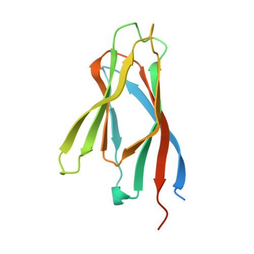
    Nucleophosmin (NPM1) is a multifunctional phospho-protein with critical roles in ribosome biogenesis, tumor suppression, and nucleolar stress response. Here we show that the N-terminal oligomerization domain of NPM1 (Npm-N) exhibits structural polymorphism by populating conformational states ranging from a highly ordered, folded pentamer to a highly disordered monomer. The monomer-pentamer equilibrium is modulated by posttranslational modification and protein binding. Phosphorylation drives the equilibrium in favor of monomeric forms, and this effect can be reversed by Npm-N binding to its interaction partners. We have identified a short, arginine-rich linear motif in NPM1 binding partners that mediates Npm-N oligomerization. We propose that the diverse functional repertoire associated with NPM1 is controlled through a regulated unfolding mechanism signaled through posttranslational modifications and intermolecular interactions.


  • Organizational Affiliation

    Department of Structural Biology, St. Jude Children's Research Hospital, Memphis, TN 38105.


Macromolecules
Find similar proteins by:  (by identity cutoff)  |  3D Structure
Entity ID: 1
MoleculeChains Sequence LengthOrganismDetailsImage
Nucleophosmin
A, B, C, D, E
133Mus musculusMutation(s): 0 
Gene Names: Npm1
UniProt
Find proteins for Q61937 (Mus musculus)
Explore Q61937 
Go to UniProtKB:  Q61937
Entity Groups  
Sequence Clusters30% Identity50% Identity70% Identity90% Identity95% Identity100% Identity
UniProt GroupQ61937
Sequence Annotations
Expand
  • Reference Sequence
Experimental Data & Validation

Experimental Data

  • Method: X-RAY DIFFRACTION
  • Resolution: 1.80 Å
  • R-Value Free: 0.190 
  • R-Value Work: 0.163 
  • R-Value Observed: 0.165 
  • Space Group: C 1 2 1
Unit Cell:
Length ( Å )Angle ( ˚ )
a = 90.675α = 90
b = 69.794β = 99.87
c = 88.63γ = 90
Software Package:
Software NamePurpose
SERGUIdata collection
PHASERphasing
PHENIXrefinement
HKL-2000data reduction
HKL-2000data scaling

Structure Validation

View Full Validation Report


Entry History 

Deposition Data

Revision History  (Full details and data files)

  • Version 1.0: 2014-03-12
    Type: Initial release
  • Version 1.1: 2014-04-23
    Changes: Database references
  • Version 1.2: 2019-11-20
    Changes: Database references, Derived calculations
  • Version 1.3: 2023-09-20
    Changes: Data collection, Database references, Derived calculations, Refinement description