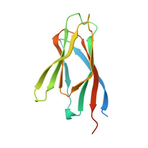
4N8M | pdb_00004n8m

Structural polymorphism in the N-terminal oligomerization domain of NPM1


Experimental Data Snapshot

  • Method: X-RAY DIFFRACTION
  • Resolution: 1.80 Å
  • R-Value Free: 
    0.190 (Depositor), 0.186 (DCC) 
  • R-Value Work: 
    0.163 (Depositor), 0.159 (DCC) 
  • R-Value Observed: 
    0.165 (Depositor) 

Starting Model: experimental
View more details

wwPDB Validation 3D Report Full Report

Validation slider image for 4N8M

This is version 1.3 of the entry. See complete history. 

Literature

Structural polymorphism in the N-terminal oligomerization domain of NPM1.

Mitrea, D.M., Grace, C.R., Buljan, M., Yun, M.K., Pytel, N.J., Satumba, J., Nourse, A., Park, C.G., Madan Babu, M., White, S.W., Kriwacki, R.W.

(2014) Proc Natl Acad Sci U S A 111: 4466-4471

  • DOI: https://doi.org/10.1073/pnas.1321007111
  • Primary Citation Related Structures: 
    4N8M

  • PubMed Abstract: 

    Nucleophosmin (NPM1) is a multifunctional phospho-protein with critical roles in ribosome biogenesis, tumor suppression, and nucleolar stress response. Here we show that the N-terminal oligomerization domain of NPM1 (Npm-N) exhibits structural polymorphism by populating conformational states ranging from a highly ordered, folded pentamer to a highly disordered monomer. The monomer-pentamer equilibrium is modulated by posttranslational modification and protein binding. Phosphorylation drives the equilibrium in favor of monomeric forms, and this effect can be reversed by Npm-N binding to its interaction partners. We have identified a short, arginine-rich linear motif in NPM1 binding partners that mediates Npm-N oligomerization. We propose that the diverse functional repertoire associated with NPM1 is controlled through a regulated unfolding mechanism signaled through posttranslational modifications and intermolecular interactions.


  • Organizational Affiliation: 
    • Department of Structural Biology, St. Jude Children's Research Hospital, Memphis, TN 38105.

Macromolecule Content 

  • Total Structure Weight: 73.37 kDa 
  • Atom Count: 4,263 
  • Modeled Residue Count: 532 
  • Deposited Residue Count: 665 
  • Unique protein chains: 1

Macromolecules

Find similar proteins by:|  3D Structure
Entity ID: 1
MoleculeChains  Sequence LengthOrganismDetailsImage
Nucleophosmin
A, B, C, D, E
133Mus musculusMutation(s): 0 
Gene Names: Npm1
UniProt
Find proteins for Q61937 (Mus musculus)
Explore Q61937 
Go to UniProtKB:  Q61937
Entity Groups
Sequence Clusters30% Identity50% Identity70% Identity90% Identity95% Identity100% Identity
UniProt GroupQ61937
Sequence Annotations
Expand
Reference Sequence

Experimental Data & Validation

Experimental Data

  • Method: X-RAY DIFFRACTION
  • Resolution: 1.80 Å
  • R-Value Free:  0.190 (Depositor), 0.186 (DCC) 
  • R-Value Work:  0.163 (Depositor), 0.159 (DCC) 
  • R-Value Observed: 0.165 (Depositor) 
Space Group: C 1 2 1
Unit Cell:
Length ( Å )Angle ( ˚ )
a = 90.675α = 90
b = 69.794β = 99.87
c = 88.63γ = 90
Software Package:
Software NamePurpose
SERGUIdata collection
PHASERphasing
PHENIXrefinement
HKL-2000data reduction
HKL-2000data scaling

Structure Validation

View Full Validation Report


Entry History 

Deposition Data

Revision History  (Full details and data files)

  • Version 1.0: 2014-03-12
    Type: Initial release
  • Version 1.1: 2014-04-23
    Changes: Database references
  • Version 1.2: 2019-11-20
    Changes: Database references, Derived calculations
  • Version 1.3: 2023-09-20
    Changes: Data collection, Database references, Derived calculations, Refinement description