3WN5

Crystal structure of asymmetrically engineered Fc variant in complex with FcgRIIIa


Experimental Data Snapshot

  • Method: X-RAY DIFFRACTION
  • Resolution: 2.78 Å
  • R-Value Free: 0.274 
  • R-Value Work: 0.236 
  • R-Value Observed: 0.238 

wwPDB Validation   3D Report Full Report


Ligand Structure Quality Assessment 


This is version 2.0 of the entry. See complete history


Literature

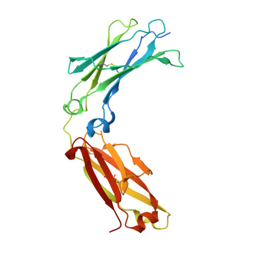
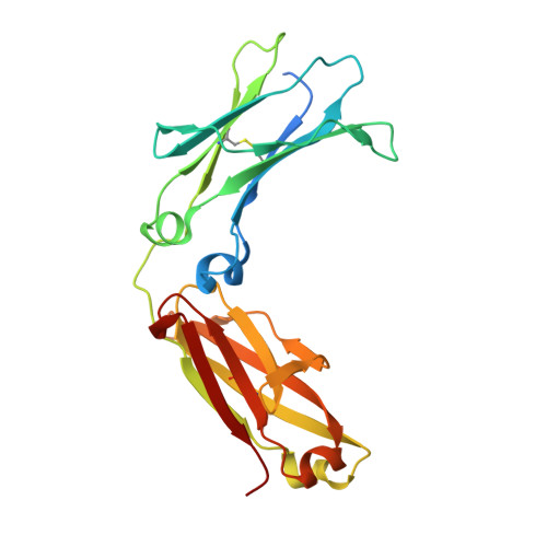
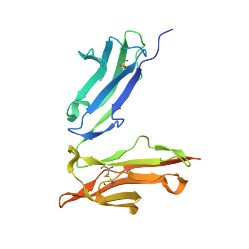
Crystal structure of a novel asymmetrically engineered Fc variant with improved affinity for Fc gamma Rs.

Mimoto, F.Kadono, S.Katada, H.Igawa, T.Kamikawa, T.Hattori, K.

(2014) Mol Immunol 58: 132-138

  • DOI: https://doi.org/10.1016/j.molimm.2013.11.017
  • Primary Citation of Related Structures:  
    3WN5

  • PubMed Abstract: 

    Enhancing the effector function by optimizing the interaction between Fc and Fcγ receptor (FcγR) is a promising approach to enhance the potency of anticancer monoclonal antibodies (mAbs). To date, a variety of Fc engineering approaches to modulate the interaction have been reported, such as afucosylation in the heavy chain Fc region or symmetrically introducing amino acid substitutions into the region, and there is still room to improve FcγR binding and thermal stability of the CH2 domain with these approaches. Recently, we have reported that asymmetric Fc engineering, which introduces different substitutions into each Fc region of heavy chain, can further improve the FcγR binding while maintaining the thermal stability of the CH2 domain by fine-tuning the asymmetric interface between the Fc domain and FcγR. However, the structural mechanism by which the asymmetrically engineered Fc improved FcγR binding remained unclear. In order to elucidate the mechanism, we solved the crystal structure of a novel asymmetrically engineered Fc, asym-mAb23, in complex with FcγRIIIa. Asym-mAb23 has enhanced binding affinity for both FcγRIIIa and FcγRIIa at the highest level of previously reported Fc variants. The structural analysis reveals the features of the asymmetrically engineered Fc in comparison with symmetric Fc and how each asymmetrically introduced substitution contributes to the improved interaction between asym-mAb23 and FcγRIIIa. This crystal structure could be utilized to enable us to design a more potent asymmetric Fc.


  • Organizational Affiliation

    Research Division, Chugai Pharmaceutical Co., Ltd., Japan.


Macromolecules
Find similar proteins by:  (by identity cutoff)  |  3D Structure
Entity ID: 1
MoleculeChains Sequence LengthOrganismDetailsImage
Ig gamma-1 chain C region
A, D
230Homo sapiensMutation(s): 9 
Gene Names: IGHG1
UniProt & NIH Common Fund Data Resources
Find proteins for P01857 (Homo sapiens)
Explore P01857 
Go to UniProtKB:  P01857
PHAROS:  P01857
GTEx:  ENSG00000211896 
Entity Groups  
Sequence Clusters30% Identity50% Identity70% Identity90% Identity95% Identity100% Identity
UniProt GroupP01857
Sequence Annotations
Expand
  • Reference Sequence
Find similar proteins by:  (by identity cutoff)  |  3D Structure
Entity ID: 2
MoleculeChains Sequence LengthOrganismDetailsImage
Ig gamma-1 chain C region
B, E
230Homo sapiensMutation(s): 10 
Gene Names: IGHG1
UniProt & NIH Common Fund Data Resources
Find proteins for P01857 (Homo sapiens)
Explore P01857 
Go to UniProtKB:  P01857
PHAROS:  P01857
GTEx:  ENSG00000211896 
Entity Groups  
Sequence Clusters30% Identity50% Identity70% Identity90% Identity95% Identity100% Identity
UniProt GroupP01857
Sequence Annotations
Expand
  • Reference Sequence
Find similar proteins by:  (by identity cutoff)  |  3D Structure
Entity ID: 3
MoleculeChains Sequence LengthOrganismDetailsImage
Low affinity immunoglobulin gamma Fc region receptor III-A
C, F
197Homo sapiensMutation(s): 3 
Gene Names: FCGR3ACD16AFCG3FCGR3IGFR3
UniProt & NIH Common Fund Data Resources
Find proteins for P08637 (Homo sapiens)
Explore P08637 
Go to UniProtKB:  P08637
PHAROS:  P08637
GTEx:  ENSG00000203747 
Entity Groups  
Sequence Clusters30% Identity50% Identity70% Identity90% Identity95% Identity100% Identity
UniProt GroupP08637
Sequence Annotations
Expand
  • Reference Sequence
Oligosaccharides

Help

Entity ID: 4
MoleculeChains Length2D Diagram Glycosylation3D Interactions
beta-D-galactopyranose-(1-4)-2-acetamido-2-deoxy-beta-D-glucopyranose-(1-2)-alpha-D-mannopyranose-(1-6)-[2-acetamido-2-deoxy-beta-D-glucopyranose-(1-2)-alpha-D-mannopyranose-(1-3)]beta-D-mannopyranose-(1-4)-2-acetamido-2-deoxy-beta-D-glucopyranose-(1-4)-[beta-L-fucopyranose-(1-6)]2-acetamido-2-deoxy-beta-D-glucopyranose
G, H, L
9N-Glycosylation
Glycosylation Resources
GlyTouCan:  G33244HE
GlyCosmos:  G33244HE
GlyGen:  G33244HE
Entity ID: 5
MoleculeChains Length2D Diagram Glycosylation3D Interactions
alpha-D-mannopyranose-(1-3)-[alpha-D-mannopyranose-(1-6)]beta-D-mannopyranose-(1-4)-2-acetamido-2-deoxy-beta-D-glucopyranose-(1-4)-2-acetamido-2-deoxy-beta-D-glucopyranose
I
5N-Glycosylation
Glycosylation Resources
GlyTouCan:  G22768VO
GlyCosmos:  G22768VO
GlyGen:  G22768VO
Entity ID: 6
MoleculeChains Length2D Diagram Glycosylation3D Interactions
2-acetamido-2-deoxy-beta-D-glucopyranose-(1-4)-2-acetamido-2-deoxy-beta-D-glucopyranose
J, M
2N-Glycosylation
Glycosylation Resources
GlyTouCan:  G42666HT
GlyCosmos:  G42666HT
GlyGen:  G42666HT
Entity ID: 7
MoleculeChains Length2D Diagram Glycosylation3D Interactions
2-acetamido-2-deoxy-beta-D-glucopyranose-(1-2)-alpha-D-mannopyranose-(1-3)-[2-acetamido-2-deoxy-beta-D-glucopyranose-(1-2)-alpha-D-mannopyranose-(1-6)]beta-D-mannopyranose-(1-4)-2-acetamido-2-deoxy-beta-D-glucopyranose-(1-4)-[beta-L-fucopyranose-(1-6)]2-acetamido-2-deoxy-beta-D-glucopyranose
K
8N-Glycosylation
Glycosylation Resources
GlyTouCan:  G59451NL
GlyCosmos:  G59451NL
GlyGen:  G59451NL
Small Molecules
Ligands 2 Unique
IDChains Name / Formula / InChI Key2D Diagram3D Interactions
NAG
Query on NAG

Download Ideal Coordinates CCD File 
Z [auth F]2-acetamido-2-deoxy-beta-D-glucopyranose
C8 H15 N O6
OVRNDRQMDRJTHS-FMDGEEDCSA-N
IOD
Query on IOD

Download Ideal Coordinates CCD File 
AA [auth F]
BA [auth F]
CA [auth F]
DA [auth F]
N [auth A]
AA [auth F],
BA [auth F],
CA [auth F],
DA [auth F],
N [auth A],
O [auth A],
P [auth B],
Q [auth B],
R [auth B],
S [auth C],
T [auth C],
U [auth D],
V [auth E],
W [auth E],
X [auth E],
Y [auth E]
IODIDE ION
I
XMBWDFGMSWQBCA-UHFFFAOYSA-M
Experimental Data & Validation

Experimental Data

  • Method: X-RAY DIFFRACTION
  • Resolution: 2.78 Å
  • R-Value Free: 0.274 
  • R-Value Work: 0.236 
  • R-Value Observed: 0.238 
  • Space Group: P 1 21 1
Unit Cell:
Length ( Å )Angle ( ˚ )
a = 75.03α = 90
b = 72.49β = 91.15
c = 163.48γ = 90
Software Package:
Software NamePurpose
XSCALEdata scaling
REFMACrefinement
PDB_EXTRACTdata extraction
XDSdata reduction
PHASERphasing

Structure Validation

View Full Validation Report


Ligand Structure Quality Assessment 


Entry History 

Deposition Data

Revision History  (Full details and data files)

  • Version 1.0: 2014-11-19
    Type: Initial release
  • Version 2.0: 2020-07-29
    Type: Remediation
    Reason: Carbohydrate remediation
    Changes: Atomic model, Data collection, Database references, Derived calculations, Structure summary