3W30

Structual basis for the recognition of Ubc13 by the Shigella flexneri effector OspI


Experimental Data Snapshot

  • Method: X-RAY DIFFRACTION
  • Resolution: 2.99 Å
  • R-Value Free: 0.303 
  • R-Value Work: 0.230 
  • R-Value Observed: 0.233 

wwPDB Validation   3D Report Full Report


This is version 1.2 of the entry. See complete history


Literature

Structural Basis for the Recognition of Ubc13 by the Shigella flexneri Effector OspI.

Nishide, A.Kim, M.Takagi, K.Himeno, A.Sanada, T.Sasakawa, C.Mizushima, T.

(2013) J Mol Biol 425: 2623-2631

  • DOI: https://doi.org/10.1016/j.jmb.2013.02.037
  • Primary Citation of Related Structures:  
    3W30, 3W31

  • PubMed Abstract: 

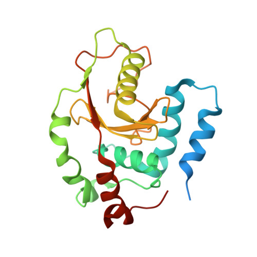
    Ubc13 is a ubiquitin-conjugating enzyme that plays a key role in the nuclear factor-κB signal transduction pathway in human diseases. The Shigella flexneri effector OspI affects inflammatory responses by catalyzing the deamidation of a specific glutamine residue at position 100 in Ubc13 during infection. This modification prevents the activation of the TNF (tumor necrosis factor) receptor-associated factor 6, leading to modulation of the diacylglycerol-CBM (CARD-Bcl10-Malt1) complex-TNF receptor-associated factor 6-nuclear factor-κB signaling pathway. To elucidate the structural basis of OspI function, we determined the crystal structures of the catalytically inert OspI C62A mutant and its complex with Ubc13 at resolutions of 3.0 and 2.96Å, respectively. The structure of the OspI-Ubc13 complex revealed that the interacting surfaces between OspI and Ubc13 are a hydrophobic surface and a complementary charged surface. Furthermore, we predict that the complementary charged surface of OspI plays a key role in substrate specificity determination.


  • Organizational Affiliation

    Picobiology Institute, Department of Life Science, Graduate School of Life Science, University of Hyogo, 3-2-1 Kouto, Kamigori-cho, Ako-gun, Hyogo 678-1297, Japan.


Macromolecules
Find similar proteins by:  (by identity cutoff)  |  3D Structure
Entity ID: 1
MoleculeChains Sequence LengthOrganismDetailsImage
ORF169b
A, B
220Shigella flexneriMutation(s): 1 
Gene Names: ORF169bCP0209
UniProt
Find proteins for Q8VSD5 (Shigella flexneri)
Explore Q8VSD5 
Go to UniProtKB:  Q8VSD5
Entity Groups  
Sequence Clusters30% Identity50% Identity70% Identity90% Identity95% Identity100% Identity
UniProt GroupQ8VSD5
Sequence Annotations
Expand
  • Reference Sequence
Experimental Data & Validation

Experimental Data

  • Method: X-RAY DIFFRACTION
  • Resolution: 2.99 Å
  • R-Value Free: 0.303 
  • R-Value Work: 0.230 
  • R-Value Observed: 0.233 
  • Space Group: P 21 21 21
Unit Cell:
Length ( Å )Angle ( ˚ )
a = 67.583α = 90
b = 67.352β = 90
c = 84.84γ = 90
Software Package:
Software NamePurpose
HKL-2000data collection
MOLREPphasing
REFMACrefinement
HKL-2000data reduction
HKL-2000data scaling

Structure Validation

View Full Validation Report


Entry History 

Deposition Data

Revision History  (Full details and data files)

  • Version 1.0: 2013-03-27
    Type: Initial release
  • Version 1.1: 2013-07-17
    Changes: Database references
  • Version 1.2: 2023-11-08
    Changes: Data collection, Database references, Refinement description