3VOW

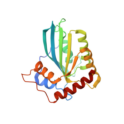
Crystal Structure of the Human APOBEC3C having HIV-1 Vif-binding Interface


Experimental Data Snapshot

  • Method: X-RAY DIFFRACTION
  • Resolution: 2.15 Å
  • R-Value Free: 0.263 
  • R-Value Work: 0.214 
  • R-Value Observed: 0.216 

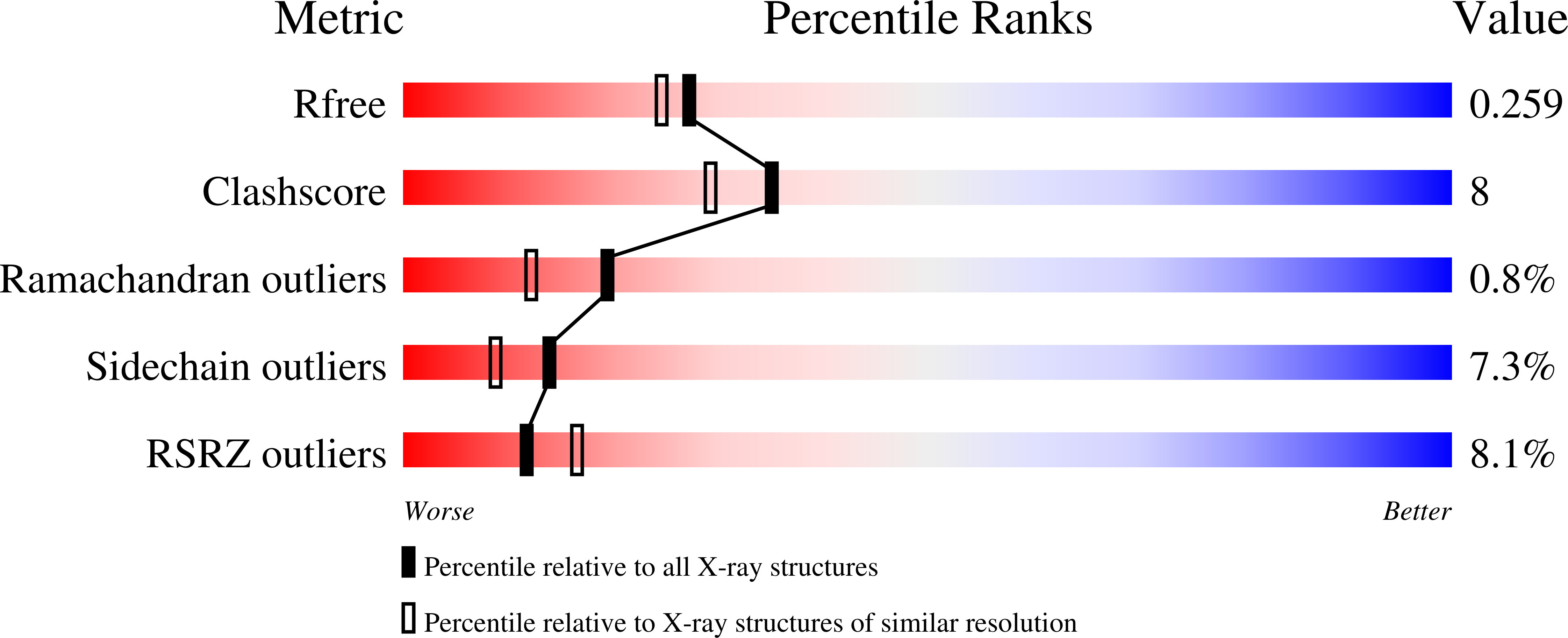
wwPDB Validation   3D Report Full Report


This is version 1.4 of the entry. See complete history


Literature

The APOBEC3C crystal structure and the interface for HIV-1 Vif binding.

Kitamura, S.Ode, H.Nakashima, M.Imahashi, M.Naganawa, Y.Kurosawa, T.Yokomaku, Y.Yamane, T.Watanabe, N.Suzuki, A.Sugiura, W.Iwatani, Y.

(2012) Nat Struct Mol Biol 19: 1005-1010

  • DOI: https://doi.org/10.1038/nsmb.2378
  • Primary Citation of Related Structures:  
    3VOW

  • PubMed Abstract: 

    The human apolipoprotein B mRNA-editing enzyme catalytic polypeptide-like 3 (APOBEC3, referred to as A3) proteins are cellular cytidine deaminases that potently restrict retrovirus replication. However, HIV-1 viral infectivity factor (Vif) counteracts the antiviral activity of most A3 proteins by targeting them for proteasomal degradation. To date, the structure of an A3 protein containing a Vif-binding interface has not been solved. Here, we report a high-resolution crystal structure of APOBEC3C and identify the HIV-1 Vif-interaction interface. Extensive structure-guided mutagenesis revealed the role of a shallow cavity composed of hydrophobic or negatively charged residues between the α2 and α3 helices. This region is distant from the DPD motif (residues 128-130) of APOBEC3G that participates in HIV-1 Vif interaction. These findings provide insight into Vif-A3 interactions and could lead to the development of new pharmacologic anti-HIV-1 compounds.


  • Organizational Affiliation

    Clinical Research Center, Department of Infectious Diseases and Immunology, National Hospital Organization Nagoya Medical Center, Nagoya, Japan.


Macromolecules
Find similar proteins by:  (by identity cutoff)  |  3D Structure
Entity ID: 1
MoleculeChains Sequence LengthOrganismDetailsImage
Probable DNA dC->dU-editing enzyme APOBEC-3C
A, B
190Homo sapiensMutation(s): 0 
Gene Names: APOBEC3CAPOBEC1LPBI
EC: 3.5.4
UniProt & NIH Common Fund Data Resources
Find proteins for Q9NRW3 (Homo sapiens)
Explore Q9NRW3 
Go to UniProtKB:  Q9NRW3
PHAROS:  Q9NRW3
GTEx:  ENSG00000244509 
Entity Groups  
Sequence Clusters30% Identity50% Identity70% Identity90% Identity95% Identity100% Identity
UniProt GroupQ9NRW3
Sequence Annotations
Expand
  • Reference Sequence
Experimental Data & Validation

Experimental Data

  • Method: X-RAY DIFFRACTION
  • Resolution: 2.15 Å
  • R-Value Free: 0.263 
  • R-Value Work: 0.214 
  • R-Value Observed: 0.216 
  • Space Group: P 61
Unit Cell:
Length ( Å )Angle ( ˚ )
a = 105.042α = 90
b = 105.042β = 90
c = 70.046γ = 120
Software Package:
Software NamePurpose
DENZOdata reduction
SCALEPACKdata scaling
MOLREPphasing
REFMACrefinement
PDB_EXTRACTdata extraction
ADSCdata collection
HKL-2000data reduction
HKL-2000data scaling

Structure Validation

View Full Validation Report


Entry History 

Deposition Data

Revision History  (Full details and data files)

  • Version 1.0: 2012-10-03
    Type: Initial release
  • Version 1.1: 2012-10-17
    Changes: Database references
  • Version 1.2: 2012-10-24
    Changes: Structure summary
  • Version 1.3: 2017-11-22
    Changes: Refinement description
  • Version 1.4: 2023-11-08
    Changes: Data collection, Database references, Derived calculations, Refinement description