3V6Q

Crystal structure of the complex of bovine lactoperoxidase with Carbon monoxide at 2.0 A resolution


Experimental Data Snapshot

  • Method: X-RAY DIFFRACTION
  • Resolution: 2.00 Å
  • R-Value Free: 0.248 
  • R-Value Work: 0.191 
  • R-Value Observed: 0.194 

wwPDB Validation   3D Report Full Report


Ligand Structure Quality Assessment 


This is version 2.2 of the entry. See complete history


Literature

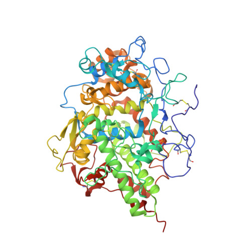
Bovine carbonyl lactoperoxidase structure at 2.0 angstrom resolution and infrared spectra as a function of pH.

Singh, A.K.Smith, M.L.Yamini, S.Ohlsson, P.I.Sinha, M.Kaur, P.Sharma, S.Paul, J.A.Singh, T.P.Paul, K.G.

(2012) Protein J 31: 598-608

  • DOI: https://doi.org/10.1007/s10930-012-9436-3
  • Primary Citation of Related Structures:  
    3V6Q

  • PubMed Abstract: 

    Lactoperoxidase (LPO) is a hemeprotein catalyzing the oxidation of thiocyanate and I(-) into antimicrobials and small aromatic organics after being itself oxidized by H(2)O(2). LPO is excreted by the lungs, mammary glands, found in saliva and tears and protects mammals against bacterial, fungal and viral invasion. The Fe(II) form binds CO which inactivates LPO like many other hemeproteins. We present the 3-dimensional structure of CO-LPO at 2.0Å resolution and infrared (IR) spectra of the iron-bound CO stretch from pH 3 to 8.8 at 1 cm(-1) resolution. The observed Fe-C-O bond angle of 132° is more acute than the electronically related Fe(III), CN-LPO with a Fe-C-N angle of 161°. The orientations of the two ligands are different with the oxygen of CO pointing towards the imidazole of distal His109 while the nitrogen of CN points away, the Fe(II) moves towards His109 while the Fe(III) moves away; both movements are consistent with a hydrogen bond between the distal His109 and CO, but not to the nitrogen of CN-LPO. The IR spectra of CO-LPO exhibit two major CO absorbances with pH dependent relative intensities. Both crystallographic and IR data suggest proton donation to the CO oxygen by His109 with a pK ≈ 4; close to the pH of greatest enzyme turnover. The IR absorbance maxima are consistent with a first order correlation between frequency and Fe(III)/Fe(II) reduction potential at pH 7; both band widths at half-height correlate with electron density donation from Fe(II) to CO as gauged by the reduction potential.


  • Organizational Affiliation

    Department of Biophysics, All India Institute of Medical Sciences, New Delhi, 110 029, India. aksaiims@gmail.com


Macromolecules
Find similar proteins by:  (by identity cutoff)  |  3D Structure
Entity ID: 1
MoleculeChains Sequence LengthOrganismDetailsImage
Lactoperoxidase595Bos taurusMutation(s): 0 
EC: 1.11.1.7
UniProt
Find proteins for P80025 (Bos taurus)
Explore P80025 
Go to UniProtKB:  P80025
Entity Groups  
Sequence Clusters30% Identity50% Identity70% Identity90% Identity95% Identity100% Identity
UniProt GroupP80025
Sequence Annotations
Expand
  • Reference Sequence
Oligosaccharides

Help

Entity ID: 2
MoleculeChains Length2D Diagram Glycosylation3D Interactions
2-acetamido-2-deoxy-beta-D-glucopyranose-(1-4)-2-acetamido-2-deoxy-beta-D-glucopyranose
B
2N-Glycosylation
Glycosylation Resources
GlyTouCan:  G42666HT
GlyCosmos:  G42666HT
GlyGen:  G42666HT
Small Molecules
Ligands 9 Unique
IDChains Name / Formula / InChI Key2D Diagram3D Interactions
HEM
Query on HEM

Download Ideal Coordinates CCD File 
C [auth A]PROTOPORPHYRIN IX CONTAINING FE
C34 H32 Fe N4 O4
KABFMIBPWCXCRK-RGGAHWMASA-L
NAG
Query on NAG

Download Ideal Coordinates CCD File 
E [auth A],
F [auth A]
2-acetamido-2-deoxy-beta-D-glucopyranose
C8 H15 N O6
OVRNDRQMDRJTHS-FMDGEEDCSA-N
IOD
Query on IOD

Download Ideal Coordinates CCD File 
G [auth A]
H [auth A]
I [auth A]
J [auth A]
K [auth A]
G [auth A],
H [auth A],
I [auth A],
J [auth A],
K [auth A],
L [auth A],
M [auth A],
N [auth A],
O [auth A]
IODIDE ION
I
XMBWDFGMSWQBCA-UHFFFAOYSA-M
MPD
Query on MPD

Download Ideal Coordinates CCD File 
Q [auth A](4S)-2-METHYL-2,4-PENTANEDIOL
C6 H14 O2
SVTBMSDMJJWYQN-YFKPBYRVSA-N
BR
Query on BR

Download Ideal Coordinates CCD File 
P [auth A]BROMIDE ION
Br
CPELXLSAUQHCOX-UHFFFAOYSA-M
EDO
Query on EDO

Download Ideal Coordinates CCD File 
R [auth A],
S [auth A]
1,2-ETHANEDIOL
C2 H6 O2
LYCAIKOWRPUZTN-UHFFFAOYSA-N
SCN
Query on SCN

Download Ideal Coordinates CCD File 
T [auth A]THIOCYANATE ION
C N S
ZMZDMBWJUHKJPS-UHFFFAOYSA-M
CA
Query on CA

Download Ideal Coordinates CCD File 
D [auth A]CALCIUM ION
Ca
BHPQYMZQTOCNFJ-UHFFFAOYSA-N
CMO
Query on CMO

Download Ideal Coordinates CCD File 
U [auth A]CARBON MONOXIDE
C O
UGFAIRIUMAVXCW-UHFFFAOYSA-N
Modified Residues  1 Unique
IDChains TypeFormula2D DiagramParent
SEP
Query on SEP
A
L-PEPTIDE LINKINGC3 H8 N O6 PSER
Experimental Data & Validation

Experimental Data

  • Method: X-RAY DIFFRACTION
  • Resolution: 2.00 Å
  • R-Value Free: 0.248 
  • R-Value Work: 0.191 
  • R-Value Observed: 0.194 
  • Space Group: P 1 21 1
Unit Cell:
Length ( Å )Angle ( ˚ )
a = 54.3α = 90
b = 80.004β = 103.02
c = 76.551γ = 90
Software Package:
Software NamePurpose
HKL-2000data collection
AMoREphasing
REFMACrefinement
DENZOdata reduction
SCALEPACKdata scaling

Structure Validation

View Full Validation Report


Ligand Structure Quality Assessment 


Entry History 

Deposition Data

Revision History  (Full details and data files)

  • Version 1.0: 2012-02-15
    Type: Initial release
  • Version 2.0: 2020-07-29
    Type: Remediation
    Reason: Carbohydrate remediation
    Changes: Advisory, Atomic model, Data collection, Derived calculations, Structure summary
  • Version 2.1: 2023-07-19
    Changes: Database references, Structure summary
  • Version 2.2: 2023-11-08
    Changes: Data collection, Refinement description