3V0R

Crystal structure of Alternaria alternata allergen Alt a 1


Experimental Data Snapshot

  • Method: X-RAY DIFFRACTION
  • Resolution: 1.90 Å
  • R-Value Free: 0.190 
  • R-Value Work: 0.167 
  • R-Value Observed: 0.168 

wwPDB Validation   3D Report Full Report


Ligand Structure Quality Assessment 


This is version 1.2 of the entry. See complete history


Literature

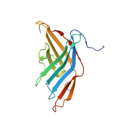
Alternaria alternata allergen Alt a 1: a unique beta-barrel protein dimer found exclusively in fungi.

Chruszcz, M.Chapman, M.D.Osinski, T.Solberg, R.Demas, M.Porebski, P.J.Majorek, K.A.Pomes, A.Minor, W.

(2012) J Allergy Clin Immunol 130: 241-7.e9

  • DOI: https://doi.org/10.1016/j.jaci.2012.03.047
  • Primary Citation of Related Structures:  
    3V0R

  • PubMed Abstract: 

    Alternaria species is one of the most common molds associated with allergic diseases, and 80% of Alternaria species-sensitive patients produce IgE antibodies to a major protein allergen, Alt a 1. The structure and function of Alt a 1 is unknown. We sought to obtain a high-resolution structure of Alt a 1 using x-ray crystallography and to investigate structural relationships between Alt a 1 and other allergens and proteins reported in the Protein Data Bank. X-ray crystallography was used to determine the structure of Alt a 1 by using a custom-designed set of crystallization conditions. An initial Alt a 1 model was determined by the application of a Ta(6)Br(12)(2+) cluster and single-wavelength anomalous diffraction. Bioinformatic analyses were used to compare the Alt a 1 sequence and structure with that of other proteins. Alt a 1 is a unique β-barrel comprising 11 β-strands and forms a "butterfly-like" dimer linked by a single disulfide bond with a large (1345 Å(2)) dimer interface. Intramolecular disulfide bonds are conserved among Alt a 1 homologs. Currently, the Alt a 1 structure has no equivalent in the Protein Data Bank. Bioinformatics analyses suggest that the structure is found exclusively in fungi. Four previously reported putative IgE-binding peptides have been located on the Alt a 1 structure. Alt a 1 has a unique, dimeric β-barrel structure that appears to define a new protein family with unknown function found exclusively in fungi. The location of IgE antibody-binding epitopes is in agreement with the structural analysis of Alt a 1. The Alt a 1 structure will allow mechanistic structure/function studies and immunologic studies directed toward new forms of immunotherapy for Alternaria species-sensitive allergic patients.


  • Organizational Affiliation

    Department of Molecular Physiology and Biological Physics, University of Virginia, Charlottesville, VA 22908, USA. maks@iwonka.med.virginia.edu


Macromolecules
Find similar proteins by:  (by identity cutoff)  |  3D Structure
Entity ID: 1
MoleculeChains Sequence LengthOrganismDetailsImage
Major allergen Alt a 1133Alternaria alternataMutation(s): 0 
Gene Names: ALTA1
UniProt
Find proteins for P79085 (Alternaria alternata)
Explore P79085 
Go to UniProtKB:  P79085
Entity Groups  
Sequence Clusters30% Identity50% Identity70% Identity90% Identity95% Identity100% Identity
UniProt GroupP79085
Sequence Annotations
Expand
  • Reference Sequence
Small Molecules
Ligands 4 Unique
IDChains Name / Formula / InChI Key2D Diagram3D Interactions
8AC
Query on 8AC

Download Ideal Coordinates CCD File 
L [auth A]8-aminooctanoic acid
C8 H17 N O2
UQXNEWQGGVUVQA-UHFFFAOYSA-N
0FU
Query on 0FU

Download Ideal Coordinates CCD File 
J [auth A],
K [auth A],
M [auth A]
2,5,6-triaminopyrimidin-4-ol
C4 H7 N5 O
SYEYEGBZVSWYPK-UHFFFAOYSA-N
SO4
Query on SO4

Download Ideal Coordinates CCD File 
B [auth A],
C [auth A],
D [auth A],
E [auth A]
SULFATE ION
O4 S
QAOWNCQODCNURD-UHFFFAOYSA-L
UNX
Query on UNX

Download Ideal Coordinates CCD File 
F [auth A],
G [auth A],
H [auth A],
I [auth A]
UNKNOWN ATOM OR ION
X
Experimental Data & Validation

Experimental Data

  • Method: X-RAY DIFFRACTION
  • Resolution: 1.90 Å
  • R-Value Free: 0.190 
  • R-Value Work: 0.167 
  • R-Value Observed: 0.168 
  • Space Group: I 41 2 2
Unit Cell:
Length ( Å )Angle ( ˚ )
a = 70.016α = 90
b = 70.016β = 90
c = 179.27γ = 90
Software Package:
Software NamePurpose
HKL-3000data collection
MD2data collection
HKL-3000phasing
MLPHAREphasing
DMmodel building
PARROTphasing
RESOLVEmodel building
ARP/wARPmodel building
CCP4model building
REFMACrefinement
Cootmodel building
HKL-2000data reduction
HKL-2000data scaling
DMphasing
RESOLVEphasing
CCP4phasing

Structure Validation

View Full Validation Report


Ligand Structure Quality Assessment 


Entry History 

Deposition Data

Revision History  (Full details and data files)

  • Version 1.0: 2012-06-13
    Type: Initial release
  • Version 1.1: 2013-06-19
    Changes: Database references
  • Version 1.2: 2022-04-13
    Changes: Database references, Derived calculations, Structure summary