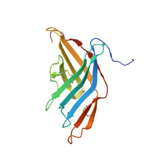
3V0R | pdb_00003v0r

Crystal structure of Alternaria alternata allergen Alt a 1


Experimental Data Snapshot

  • Method: X-RAY DIFFRACTION
  • Resolution: 1.90 Å
  • R-Value Free: 
    0.190 (Depositor), 0.200 (DCC) 
  • R-Value Work: 
    0.167 (Depositor), 0.180 (DCC) 
  • R-Value Observed: 
    0.168 (Depositor) 

wwPDB Validation   3D Report Full Report


Ligand Structure Quality Assessment 

Created with Raphaël 2.3.0Worse 01 BetterLigand structure goodness of fit to experimental dataBest fitted 8ACClick on this verticalbar to view details

This is version 1.3 of the entry. See complete history


Literature

Alternaria alternata allergen Alt a 1: a unique beta-barrel protein dimer found exclusively in fungi.

Chruszcz, M.Chapman, M.D.Osinski, T.Solberg, R.Demas, M.Porebski, P.J.Majorek, K.A.Pomes, A.Minor, W.

(2012) J Allergy Clin Immunol 130: 241-7.e9

  • DOI: https://doi.org/10.1016/j.jaci.2012.03.047
  • Primary Citation of Related Structures:  
    3V0R

  • PubMed Abstract: 

    Alternaria species is one of the most common molds associated with allergic diseases, and 80% of Alternaria species-sensitive patients produce IgE antibodies to a major protein allergen, Alt a 1. The structure and function of Alt a 1 is unknown. We sought to obtain a high-resolution structure of Alt a 1 using x-ray crystallography and to investigate structural relationships between Alt a 1 and other allergens and proteins reported in the Protein Data Bank. X-ray crystallography was used to determine the structure of Alt a 1 by using a custom-designed set of crystallization conditions. An initial Alt a 1 model was determined by the application of a Ta(6)Br(12)(2+) cluster and single-wavelength anomalous diffraction. Bioinformatic analyses were used to compare the Alt a 1 sequence and structure with that of other proteins. Alt a 1 is a unique β-barrel comprising 11 β-strands and forms a "butterfly-like" dimer linked by a single disulfide bond with a large (1345 Å(2)) dimer interface. Intramolecular disulfide bonds are conserved among Alt a 1 homologs. Currently, the Alt a 1 structure has no equivalent in the Protein Data Bank. Bioinformatics analyses suggest that the structure is found exclusively in fungi. Four previously reported putative IgE-binding peptides have been located on the Alt a 1 structure. Alt a 1 has a unique, dimeric β-barrel structure that appears to define a new protein family with unknown function found exclusively in fungi. The location of IgE antibody-binding epitopes is in agreement with the structural analysis of Alt a 1. The Alt a 1 structure will allow mechanistic structure/function studies and immunologic studies directed toward new forms of immunotherapy for Alternaria species-sensitive allergic patients.


  • Organizational Affiliation

    Department of Molecular Physiology and Biological Physics, University of Virginia, Charlottesville, VA 22908, USA. maks@iwonka.med.virginia.edu


Macromolecules
Find similar proteins by:  (by identity cutoff)  |  3D Structure
Entity ID: 1
MoleculeChains Sequence LengthOrganismDetailsImage
Major allergen Alt a 1133Alternaria alternataMutation(s): 0 
Gene Names: ALTA1
UniProt
Find proteins for P79085 (Alternaria alternata)
Explore P79085 
Go to UniProtKB:  P79085
Entity Groups  
Sequence Clusters30% Identity50% Identity70% Identity90% Identity95% Identity100% Identity
UniProt GroupP79085
Sequence Annotations
Expand
  • Reference Sequence
Small Molecules
Ligands 4 Unique
IDChains Name / Formula / InChI Key2D Diagram3D Interactions
8AC
Query on 8AC

Download Ideal Coordinates CCD File 
L [auth A]8-aminooctanoic acid
C8 H17 N O2
UQXNEWQGGVUVQA-UHFFFAOYSA-N
0FU
Query on 0FU

Download Ideal Coordinates CCD File 
J [auth A],
K [auth A],
M [auth A]
2,5,6-triaminopyrimidin-4-ol
C4 H7 N5 O
SYEYEGBZVSWYPK-UHFFFAOYSA-N
SO4
Query on SO4

Download Ideal Coordinates CCD File 
B [auth A],
C [auth A],
D [auth A],
E [auth A]
SULFATE ION
O4 S
QAOWNCQODCNURD-UHFFFAOYSA-L
UNX
Query on UNX

Download Ideal Coordinates CCD File 
F [auth A],
G [auth A],
H [auth A],
I [auth A]
UNKNOWN ATOM OR ION
X
Experimental Data & Validation

Experimental Data

  • Method: X-RAY DIFFRACTION
  • Resolution: 1.90 Å
  • R-Value Free:  0.190 (Depositor), 0.200 (DCC) 
  • R-Value Work:  0.167 (Depositor), 0.180 (DCC) 
  • R-Value Observed: 0.168 (Depositor) 
Space Group: I 41 2 2
Unit Cell:
Length ( Å )Angle ( ˚ )
a = 70.016α = 90
b = 70.016β = 90
c = 179.27γ = 90
Software Package:
Software NamePurpose
HKL-3000data collection
MD2data collection
HKL-3000phasing
MLPHAREphasing
DMmodel building
PARROTphasing
RESOLVEmodel building
ARP/wARPmodel building
CCP4model building
REFMACrefinement
Cootmodel building
HKL-2000data reduction
HKL-2000data scaling
DMphasing
RESOLVEphasing
CCP4phasing

Structure Validation

View Full Validation Report


Ligand Structure Quality Assessment 

Created with Raphaël 2.3.0Worse 01 BetterLigand structure goodness of fit to experimental dataBest fitted 8ACClick on this verticalbar to view details

Entry History 

Deposition Data

Revision History  (Full details and data files)

  • Version 1.0: 2012-06-13
    Type: Initial release
  • Version 1.1: 2013-06-19
    Changes: Database references
  • Version 1.2: 2022-04-13
    Changes: Database references, Derived calculations, Structure summary
  • Version 1.3: 2024-10-30
    Changes: Data collection, Structure summary