3UEN

Crystal structure of TopBP1 BRCT4/5 domains


Experimental Data Snapshot

  • Method: X-RAY DIFFRACTION
  • Resolution: 1.90 Å
  • R-Value Free: 0.224 
  • R-Value Work: 0.175 
  • R-Value Observed: 0.177 

wwPDB Validation   3D Report Full Report


This is version 1.3 of the entry. See complete history


Literature

Structural insights into recognition of MDC1 by TopBP1 in DNA replication checkpoint control.

Leung, C.C.Sun, L.Gong, Z.Burkat, M.Edwards, R.Assmus, M.Chen, J.Glover, J.N.

(2013) Structure 21: 1450-1459

  • DOI: https://doi.org/10.1016/j.str.2013.06.015
  • Primary Citation of Related Structures:  
    3UEN, 3UEO

  • PubMed Abstract: 

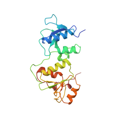
    Activation of the DNA replication checkpoint by the ATR kinase requires protein interactions mediated by the ATR-activating protein, TopBP1. Accumulation of TopBP1 at stalled replication forks requires the interaction of TopBP1 BRCT5 with the phosphorylated SDT repeats of the adaptor protein MDC1. Here, we present the X-ray crystal structures of the tandem BRCT4/5 domains of TopBP1 free and in complex with a MDC1 consensus pSDpT phosphopeptide. TopBP1 BRCT4/5 adopts a variant BRCT-BRCT packing interface and recognizes its target peptide in a manner distinct from that observed in previous tandem BRCT- peptide structures. The phosphate-binding pocket and positively charged residues in a variant loop in BRCT5 present an extended binding surface for the negatively charged MDC1 phosphopeptide. Mutations in this surface reduce binding affinity and recruitment of TopBP1 to γH2AX foci in cells. These studies reveal a different mode of phosphopeptide binding by BRCT domains in the DNA damage response.


  • Organizational Affiliation

    Department of Biochemistry, University of Alberta, Edmonton, Alberta T6G 2H7, Canada.


Macromolecules
Find similar proteins by:  (by identity cutoff)  |  3D Structure
Entity ID: 1
MoleculeChains Sequence LengthOrganismDetailsImage
DNA topoisomerase 2-binding protein 1203Homo sapiensMutation(s): 0 
Gene Names: KIAA0259TOPBP1
UniProt & NIH Common Fund Data Resources
Find proteins for Q92547 (Homo sapiens)
Explore Q92547 
Go to UniProtKB:  Q92547
PHAROS:  Q92547
GTEx:  ENSG00000163781 
Entity Groups  
Sequence Clusters30% Identity50% Identity70% Identity90% Identity95% Identity100% Identity
UniProt GroupQ92547
Sequence Annotations
Expand
  • Reference Sequence
Experimental Data & Validation

Experimental Data

  • Method: X-RAY DIFFRACTION
  • Resolution: 1.90 Å
  • R-Value Free: 0.224 
  • R-Value Work: 0.175 
  • R-Value Observed: 0.177 
  • Space Group: P 2 2 21
Unit Cell:
Length ( Å )Angle ( ˚ )
a = 35.902α = 90
b = 48.801β = 90
c = 126.088γ = 90
Software Package:
Software NamePurpose
DENZOdata reduction
SCALEPACKdata scaling
PHASERphasing
PHENIXrefinement
PDB_EXTRACTdata extraction
HKL-2000data collection
HKL-2000data reduction
HKL-2000data scaling

Structure Validation

View Full Validation Report


Entry History 

Deposition Data

Revision History  (Full details and data files)

  • Version 1.0: 2013-07-03
    Type: Initial release
  • Version 1.1: 2014-03-26
    Changes: Database references
  • Version 1.2: 2017-11-08
    Changes: Refinement description
  • Version 1.3: 2024-02-28
    Changes: Data collection, Database references, Derived calculations