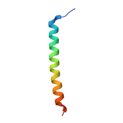
Crystal structure and characterization of coiled-coil domain of the transient receptor potential channel PKD2L1.
Molland, K.L., Paul, L.N., Yernool, D.A.(2011) Biochim Biophys Acta 1824: 413-421
- PubMed: 22193359
- DOI: https://doi.org/10.1016/j.bbapap.2011.12.002
- Primary Citation of Related Structures:
3TE3 - PubMed Abstract:
The cation-permeable channel PKD2L1 forms a homomeric assembly as well as heteromeric associations with both PKD1 and PKD1L3, with the cytoplasmic regulatory domain (CRD) of PKD2L1 often playing a role in assembly and/or function. Our previous work indicated that the isolated PKD2L1 CRD assembles as a trimer in a manner dependent on the presence of a proposed oligomerization domain. Herein we describe the 2.7Å crystal structure of a segment containing the PKD2L1 oligomerization domain which indicates that trimerization is driven by the β-branched residues at the first and fourth positions of a heptad repeat (commonly referred to as "a" and "d") and by a conserved R-h-x-x-h-E salt bridge motif that is largely unique to parallel trimeric coiled coils. Further analysis of the PKD2L1 CRD indicates that trimeric association is sufficiently strong that no other species are present in solution in an analytical ultracentrifugation experiment at the lowest measurable concentration of 750nM. Conversely, mutation of the "a" and "d" residues leads to formation of an exclusively monomeric species, independent of concentration. Although both monomeric and WT CRDs are stable in solution and bind calcium with 0.9μM affinity, circular dichroism studies reveal that the monomer loses 25% more α-helical content than WT when stripped of this ligand, suggesting that the CRD structure is stabilized by trimerization in the ligand-free state. This stability could play a role in the function of the full-length complex, indicating that trimerization may be important for both homo- and possibly heteromeric assemblies of PKD2L1.
Organizational Affiliation:
Department of Biological Sciences, Purdue University, West Lafayette, IN 47907, USA. kmolland@purdue.edu