3T1P

Crystal structure of an alpha-1-antitrypsin trimer


Experimental Data Snapshot

  • Method: X-RAY DIFFRACTION
  • Resolution: 3.90 Å
  • R-Value Free: 0.297 
  • R-Value Work: 0.231 
  • R-Value Observed: 0.238 

wwPDB Validation   3D Report Full Report


This is version 1.2 of the entry. See complete history


Literature

Molecular basis of alpha 1-antitrypsin deficiency revealed by the structure of a domain-swapped trimer.

Yamasaki, M.Sendall, T.J.Pearce, M.C.Whisstock, J.C.Huntington, J.A.

(2011) EMBO Rep 12: 1011-1017

  • DOI: https://doi.org/10.1038/embor.2011.171
  • Primary Citation of Related Structures:  
    3T1P

  • PubMed Abstract: 

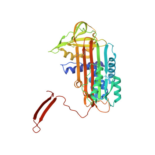
    α(1)-Antitrypsin (α1AT) deficiency is a disease with multiple manifestations, including cirrhosis and emphysema, caused by the accumulation of stable polymers of mutant protein in the endoplasmic reticulum of hepatocytes. However, the molecular basis of misfolding and polymerization remain unknown. We produced and crystallized a trimeric form of α1AT that is recognized by an antibody specific for the pathological polymer. Unexpectedly, this structure reveals a polymeric linkage mediated by domain swapping the carboxy-terminal 34 residues. Disulphide-trapping and antibody-binding studies further demonstrate that runaway C-terminal domain swapping, rather than the s4A/s5A domain swap previously proposed, underlies polymerization of the common Z-mutant of α1AT in vivo.


  • Organizational Affiliation

    Department of Haematology, Cambridge Institute for Medical Research, University of Cambridge, Wellcome Trust/MRC Building, Hills Road, Cambridge CB2 0XY, UK.


Macromolecules
Find similar proteins by:  (by identity cutoff)  |  3D Structure
Entity ID: 1
MoleculeChains Sequence LengthOrganismDetailsImage
Alpha-1-antitrypsin371Homo sapiensMutation(s): 4 
Gene Names: SERPINA1AATPIPRO0684PRO2209
UniProt & NIH Common Fund Data Resources
Find proteins for P01009 (Homo sapiens)
Explore P01009 
Go to UniProtKB:  P01009
PHAROS:  P01009
GTEx:  ENSG00000197249 
Entity Groups  
Sequence Clusters30% Identity50% Identity70% Identity90% Identity95% Identity100% Identity
UniProt GroupP01009
Sequence Annotations
Expand
  • Reference Sequence
Experimental Data & Validation

Experimental Data

  • Method: X-RAY DIFFRACTION
  • Resolution: 3.90 Å
  • R-Value Free: 0.297 
  • R-Value Work: 0.231 
  • R-Value Observed: 0.238 
  • Space Group: P 41 3 2
Unit Cell:
Length ( Å )Angle ( ˚ )
a = 163.691α = 90
b = 163.691β = 90
c = 163.691γ = 90
Software Package:
Software NamePurpose
DNAdata collection
PHASERphasing
REFMACrefinement
MOSFLMdata reduction
SCALAdata scaling

Structure Validation

View Full Validation Report


Entry History 

Deposition Data

Revision History  (Full details and data files)

  • Version 1.0: 2011-08-17
    Type: Initial release
  • Version 1.1: 2017-06-21
    Changes: Database references
  • Version 1.2: 2023-09-13
    Changes: Data collection, Database references, Refinement description