3SE4

human IFNw-IFNAR ternary complex


Experimental Data Snapshot

  • Method: X-RAY DIFFRACTION
  • Resolution: 3.50 Å
  • R-Value Free: 0.288 
  • R-Value Work: 0.213 

wwPDB Validation   3D Report Full Report


Ligand Structure Quality Assessment 


This is version 1.4 of the entry. See complete history


Literature

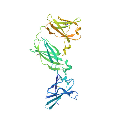
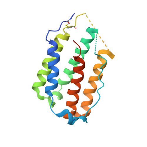
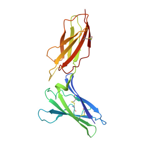
Structural linkage between ligand discrimination and receptor activation by type I interferons.

Thomas, C.Moraga, I.Levin, D.Krutzik, P.O.Podoplelova, Y.Trejo, A.Lee, C.Yarden, G.Vleck, S.E.Glenn, J.S.Nolan, G.P.Piehler, J.Schreiber, G.Garcia, K.C.

(2011) Cell 146: 621-632

  • DOI: https://doi.org/10.1016/j.cell.2011.06.048
  • Primary Citation of Related Structures:  
    3S8W, 3S98, 3S9D, 3SE3, 3SE4

  • PubMed Abstract: 

    Type I Interferons (IFNs) are important cytokines for innate immunity against viruses and cancer. Sixteen human type I IFN variants signal through the same cell-surface receptors, IFNAR1 and IFNAR2, yet they can evoke markedly different physiological effects. The crystal structures of two human type I IFN ternary signaling complexes containing IFNα2 and IFNω reveal recognition modes and heterotrimeric architectures that are unique among the cytokine receptor superfamily but conserved between different type I IFNs. Receptor-ligand cross-reactivity is enabled by conserved receptor-ligand "anchor points" interspersed among ligand-specific interactions that "tune" the relative IFN-binding affinities, in an apparent extracellular "ligand proofreading" mechanism that modulates biological activity. Functional differences between IFNs are linked to their respective receptor recognition chemistries, in concert with a ligand-induced conformational change in IFNAR1, that collectively control signal initiation and complex stability, ultimately regulating differential STAT phosphorylation profiles, receptor internalization rates, and downstream gene expression patterns.


  • Organizational Affiliation

    Howard Hughes Medical Institute, Department of Molecular and Cellular Physiology, Stanford University School of Medicine, Stanford, CA 94305, USA.


Macromolecules
Find similar proteins by:  (by identity cutoff)  |  3D Structure
Entity ID: 1
MoleculeChains Sequence LengthOrganismDetailsImage
Interferon alpha/beta receptor 1414Homo sapiensMutation(s): 0 
Gene Names: IFNAR1IFNAR
UniProt & NIH Common Fund Data Resources
Find proteins for P17181 (Homo sapiens)
Explore P17181 
Go to UniProtKB:  P17181
PHAROS:  P17181
GTEx:  ENSG00000142166 
Entity Groups  
Sequence Clusters30% Identity50% Identity70% Identity90% Identity95% Identity100% Identity
UniProt GroupP17181
Sequence Annotations
Expand
  • Reference Sequence
Find similar proteins by:  (by identity cutoff)  |  3D Structure
Entity ID: 2
MoleculeChains Sequence LengthOrganismDetailsImage
Interferon omega-1177Homo sapiensMutation(s): 1 
Gene Names: IFNW1
UniProt & NIH Common Fund Data Resources
Find proteins for P05000 (Homo sapiens)
Explore P05000 
Go to UniProtKB:  P05000
GTEx:  ENSG00000177047 
Entity Groups  
Sequence Clusters30% Identity50% Identity70% Identity90% Identity95% Identity100% Identity
UniProt GroupP05000
Sequence Annotations
Expand
  • Reference Sequence
Find similar proteins by:  (by identity cutoff)  |  3D Structure
Entity ID: 3
MoleculeChains Sequence LengthOrganismDetailsImage
Interferon alpha/beta receptor 2199Homo sapiensMutation(s): 0 
Gene Names: IFNAR2IFNABRIFNARB
UniProt & NIH Common Fund Data Resources
Find proteins for P48551 (Homo sapiens)
Explore P48551 
Go to UniProtKB:  P48551
PHAROS:  P48551
GTEx:  ENSG00000159110 
Entity Groups  
Sequence Clusters30% Identity50% Identity70% Identity90% Identity95% Identity100% Identity
UniProt GroupP48551
Sequence Annotations
Expand
  • Reference Sequence
Small Molecules
Ligands 1 Unique
IDChains Name / Formula / InChI Key2D Diagram3D Interactions
NAG
Query on NAG

Download Ideal Coordinates CCD File 
D [auth A]2-acetamido-2-deoxy-beta-D-glucopyranose
C8 H15 N O6
OVRNDRQMDRJTHS-FMDGEEDCSA-N
Experimental Data & Validation

Experimental Data

  • Method: X-RAY DIFFRACTION
  • Resolution: 3.50 Å
  • R-Value Free: 0.288 
  • R-Value Work: 0.213 
  • Space Group: P 61 2 2
Unit Cell:
Length ( Å )Angle ( ˚ )
a = 93.49α = 90
b = 93.49β = 90
c = 403.13γ = 120
Software Package:
Software NamePurpose
Blu-Icedata collection
PHASERphasing
PHENIXrefinement
XDSdata reduction
XSCALEdata scaling

Structure Validation

View Full Validation Report


Ligand Structure Quality Assessment 


Entry History 

Deposition Data

Revision History  (Full details and data files)

  • Version 1.0: 2011-08-31
    Type: Initial release
  • Version 1.1: 2012-03-21
    Changes: Database references
  • Version 1.2: 2020-07-29
    Type: Remediation
    Reason: Carbohydrate remediation
    Changes: Data collection, Database references, Derived calculations, Structure summary
  • Version 1.3: 2023-09-13
    Changes: Data collection, Database references, Refinement description, Structure summary
  • Version 1.4: 2023-12-27
    Changes: Advisory, Derived calculations