


Crystal structure of a tripeptidase (SAV1512) from STAPHYLOCOCCUS AUREUS MU50 at 2.10 A resolution
Joint Center for Structural Genomics (JCSG)To be published.
Experimental Data Snapshot
wwPDB Validation 3D Report Full Report

Entity ID: 1 | |||||
|---|---|---|---|---|---|
| Molecule | Chains | Sequence Length | Organism | Details | Image |
| tripeptidase | 396 | Staphylococcus aureus subsp. aureus Mu50 | Mutation(s): 0 Gene Names: SAV1512 | 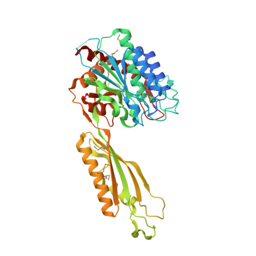 | |
UniProt | |||||
Find proteins for A0A0H3JRK2 (Staphylococcus aureus (strain Mu50 / ATCC 700699)) Explore A0A0H3JRK2 Go to UniProtKB: A0A0H3JRK2 | |||||
Entity Groups | |||||
| Sequence Clusters | 30% Identity50% Identity70% Identity90% Identity95% Identity100% Identity | ||||
| UniProt Group | A0A0H3JRK2 | ||||
Sequence AnnotationsExpand | |||||
| |||||
| Ligands 7 Unique | |||||
|---|---|---|---|---|---|
| ID | Chains | Name / Formula / InChI Key | 2D Diagram | 3D Interactions | |
| PG4 Query on PG4 | L [auth A], S [auth B], T [auth B] | TETRAETHYLENE GLYCOL C8 H18 O5 UWHCKJMYHZGTIT-UHFFFAOYSA-N |  | ||
| CIT Query on CIT | F [auth A], G [auth A] | CITRIC ACID C6 H8 O7 KRKNYBCHXYNGOX-UHFFFAOYSA-N |  | ||
| PGE Query on PGE | R [auth B] | TRIETHYLENE GLYCOL C6 H14 O4 ZIBGPFATKBEMQZ-UHFFFAOYSA-N |  | ||
| PEG Query on PEG | I [auth A], J [auth A], K [auth A], Q [auth B] | DI(HYDROXYETHYL)ETHER C4 H10 O3 MTHSVFCYNBDYFN-UHFFFAOYSA-N |  | ||
| ZN Query on ZN | C [auth A], M [auth B] | ZINC ION Zn PTFCDOFLOPIGGS-UHFFFAOYSA-N |  | ||
| IPA Query on IPA | H [auth A] | ISOPROPYL ALCOHOL C3 H8 O KFZMGEQAYNKOFK-UHFFFAOYSA-N |  | ||
| CA Query on CA | D [auth A], E [auth A], N [auth B], O [auth B], P [auth B] | CALCIUM ION Ca BHPQYMZQTOCNFJ-UHFFFAOYSA-N |  | ||
| Modified Residues 1 Unique | |||||
|---|---|---|---|---|---|
| ID | Chains | Type | Formula | 2D Diagram | Parent |
| MSE Query on MSE | A, B | L-PEPTIDE LINKING | C5 H11 N O2 Se |  | MET |
| Length ( Å ) | Angle ( ˚ ) |
|---|---|
| a = 46.996 | α = 89.67 |
| b = 57.805 | β = 73.38 |
| c = 79.857 | γ = 72.36 |
| Software Name | Purpose |
|---|---|
| MolProbity | model building |
| PDB_EXTRACT | data extraction |
| SHELX | phasing |
| SHARP | phasing |
| XSCALE | data scaling |
| REFMAC | refinement |
| XDS | data reduction |
| SHELXD | phasing |
RCSB PDB (citation) is hosted by
RCSB PDB is a member of the

