


Molecular basis of WNT Activation via the DIX-domain protein CCD1
Liu, Y.T., Dan, Q.J., Wang, J.W., Feng, Y.G., Chen, L., Liang, J., Li, Q.X., Lin, S.C., Wang, Z.X., Wu, J.W.To be published.
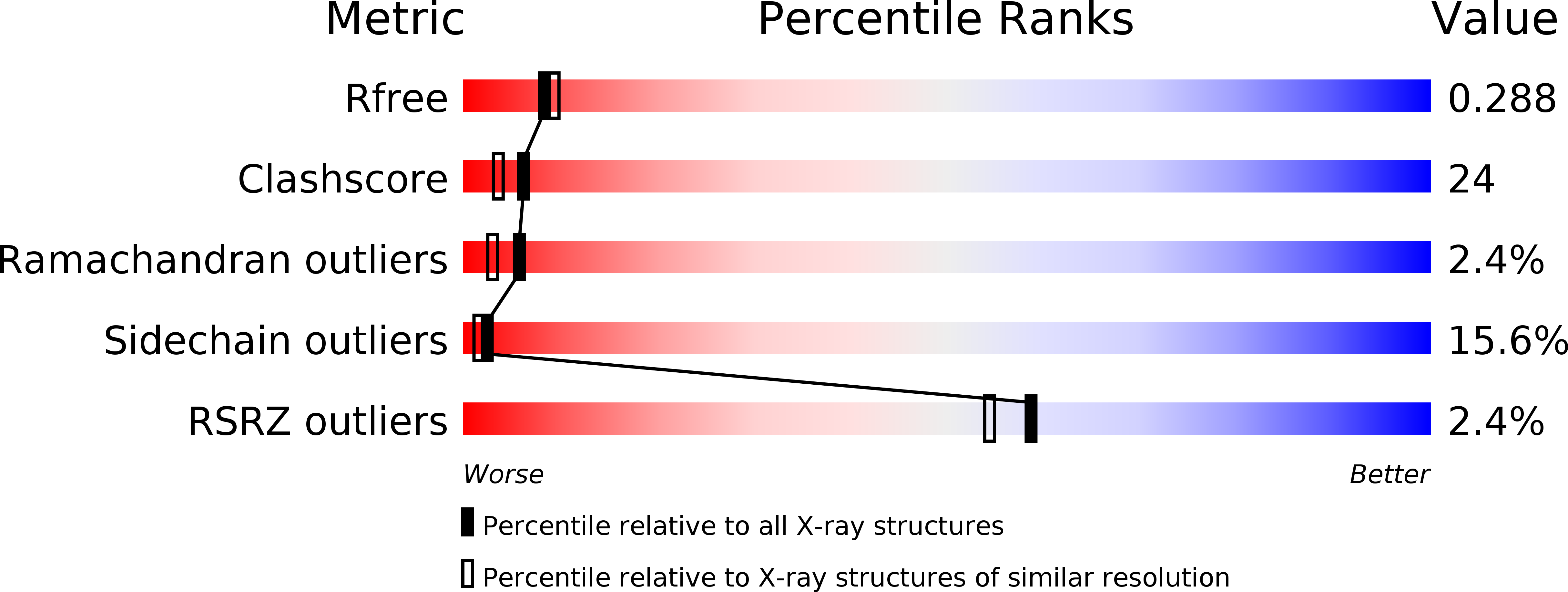
Experimental Data Snapshot
wwPDB Validation 3D Report Full Report
Entity ID: 1 | |||||
|---|---|---|---|---|---|
| Molecule | Chains | Sequence Length | Organism | Details | Image |
| Dixin | 91 | Homo sapiens | Mutation(s): 1 | 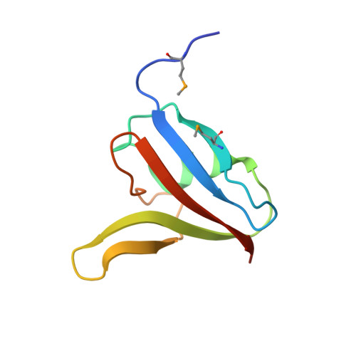 | |
UniProt & NIH Common Fund Data Resources | |||||
Find proteins for Q155Q3 (Homo sapiens) Explore Q155Q3 Go to UniProtKB: Q155Q3 | |||||
PHAROS: Q155Q3 GTEx: ENSG00000150764 | |||||
Entity Groups | |||||
| Sequence Clusters | 30% Identity50% Identity70% Identity90% Identity95% Identity100% Identity | ||||
| UniProt Group | Q155Q3 | ||||
Sequence AnnotationsExpand | |||||
| |||||
| Ligands 1 Unique | |||||
|---|---|---|---|---|---|
| ID | Chains | Name / Formula / InChI Key | 2D Diagram | 3D Interactions | |
| EDO Query on EDO | B [auth A] | 1,2-ETHANEDIOL C2 H6 O2 LYCAIKOWRPUZTN-UHFFFAOYSA-N |  | ||
| Modified Residues 1 Unique | |||||
|---|---|---|---|---|---|
| ID | Chains | Type | Formula | 2D Diagram | Parent |
| MSE Query on MSE | A | L-PEPTIDE LINKING | C5 H11 N O2 Se |  | MET |
| Length ( Å ) | Angle ( ˚ ) |
|---|---|
| a = 62.576 | α = 90 |
| b = 62.576 | β = 90 |
| c = 77.722 | γ = 120 |
| Software Name | Purpose |
|---|---|
| HKL-2000 | data collection |
| SOLVE | phasing |
| PHENIX | refinement |
| HKL-2000 | data reduction |
| HKL-2000 | data scaling |
RCSB PDB (citation) is hosted by
RCSB PDB is a member of the

