3OSP

Structure of rev1


Experimental Data Snapshot

  • Method: X-RAY DIFFRACTION
  • Resolution: 2.50 Å
  • R-Value Free: 0.255 
  • R-Value Work: 0.204 
  • R-Value Observed: 0.208 

wwPDB Validation   3D Report Full Report


Ligand Structure Quality Assessment 


This is version 1.2 of the entry. See complete history


Literature

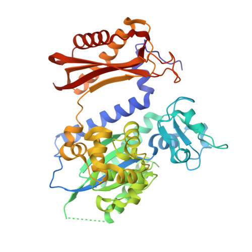
DNA synthesis across an abasic lesion by yeast REV1 DNA polymerase.

Nair, D.T.Johnson, R.E.Prakash, L.Prakash, S.Aggarwal, A.K.

(2011) J Mol Biol 406: 18-28

  • DOI: https://doi.org/10.1016/j.jmb.2010.12.016
  • Primary Citation of Related Structures:  
    3OSP

  • PubMed Abstract: 

    Abasic (apurinic/apyrimidinic) sites are among the most abundant DNA lesions in humans, and they present a strong block to replication. They are also highly mutagenic because when replicative DNA polymerases manage to insert a nucleotide opposite the lesion, they prefer to insert an A. Rev1, a member of Y-family DNA polymerases, does not obey the A-rule. This enzyme inserts a C opposite an abasic lesion with much greater catalytic efficiency than an A, G, or T. We present here the structure of yeast Rev1 in ternary complex with DNA containing an abasic lesion and with dCTP as the incoming nucleotide. The structure reveals a mechanism of synthesis across an abasic lesion that differs from that in other polymerases. The lesion is driven to an extrahelical position, and the incorporation of a C is mediated by an arginine (Arg324) that is conserved in all known orthologs of Rev1, including humans. The hydrophobic cavity that normally accommodates the unmodified G is instead filled with water molecules. Since Gs are especially prone to depurination through a spontaneous hydrolysis of the glycosidic bond, the ability of Rev1 to stabilize an abasic lesion in its active site and employ a surrogate arginine to incorporate a C provides a unique means for the "error-free" bypass of this noninstructional lesion.


  • Organizational Affiliation

    Department of Structural and Chemical Biology, Mount Sinai School of Medicine, Box 1677, 1425 Madison Avenue, New York, NY 10029, USA.


Macromolecules

Find similar proteins by:  (by identity cutoff)  |  3D Structure
Entity ID: 1
MoleculeChains Sequence LengthOrganismDetailsImage
DNA repair protein REV1434Saccharomyces cerevisiaeMutation(s): 0 
Gene Names: O6339REV1YOR346W
EC: 2.7.7
UniProt
Find proteins for P12689 (Saccharomyces cerevisiae (strain ATCC 204508 / S288c))
Explore P12689 
Go to UniProtKB:  P12689
Entity Groups  
Sequence Clusters30% Identity50% Identity70% Identity90% Identity95% Identity100% Identity
UniProt GroupP12689
Sequence Annotations
Expand
  • Reference Sequence

Find similar nucleic acids by:  Sequence   |   3D Structure  

Entity ID: 2
MoleculeChains LengthOrganismImage
5'-D(*TP*AP*AP*(3DR)P*GP*TP*AP*GP*GP*GP*GP*AP*GP*GP*AP*T)-3'B [auth T]16N/A
Sequence Annotations
Expand
  • Reference Sequence

Find similar nucleic acids by:  Sequence   |   3D Structure  

Entity ID: 3
MoleculeChains LengthOrganismImage
5'-D(*AP*TP*CP*CP*TP*CP*CP*CP*CP*TP*AP*(DOC))-3'C [auth P]12N/A
Sequence Annotations
Expand
  • Reference Sequence
Experimental Data & Validation

Experimental Data

  • Method: X-RAY DIFFRACTION
  • Resolution: 2.50 Å
  • R-Value Free: 0.255 
  • R-Value Work: 0.204 
  • R-Value Observed: 0.208 
  • Space Group: C 2 2 21
Unit Cell:
Length ( Å )Angle ( ˚ )
a = 180.021α = 90
b = 200.236β = 90
c = 55.725γ = 90
Software Package:
Software NamePurpose
Jdirectdata collection
AMoREphasing
CNSrefinement
HKL-2000data reduction
HKL-2000data scaling

Structure Validation

View Full Validation Report


Ligand Structure Quality Assessment 


Entry History 

Deposition Data

Revision History  (Full details and data files)

  • Version 1.0: 2011-04-13
    Type: Initial release
  • Version 1.1: 2011-07-13
    Changes: Version format compliance
  • Version 1.2: 2023-11-01
    Changes: Data collection, Database references, Derived calculations, Refinement description