3JC1

Electron cryo-microscopy of the IST1-CHMP1B ESCRT-III copolymer


Experimental Data Snapshot

  • Method: ELECTRON MICROSCOPY
  • Resolution: 4.00 Å
  • Aggregation State: FILAMENT 
  • Reconstruction Method: HELICAL 

wwPDB Validation   3D Report Full Report


This is version 1.3 of the entry. See complete history


Literature

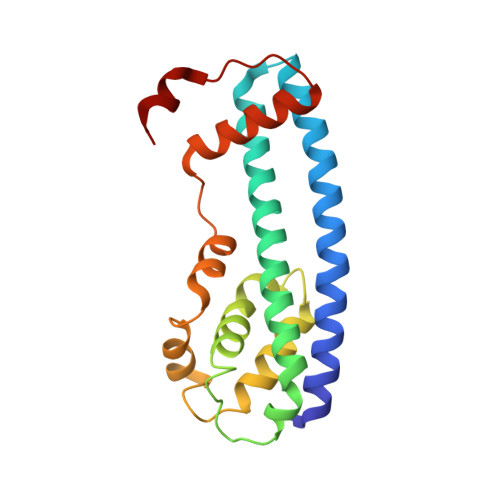
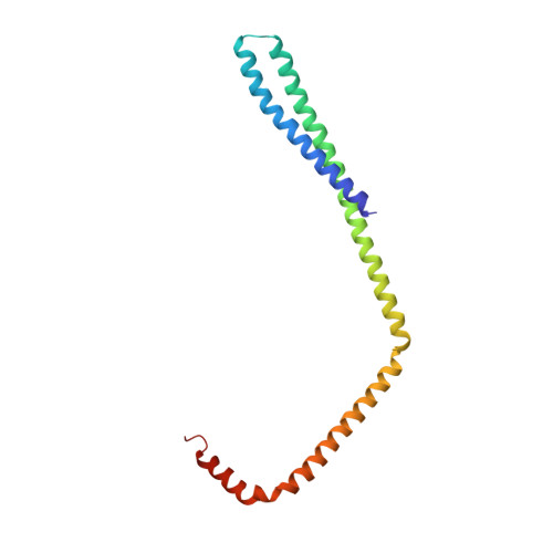
Structure and membrane remodeling activity of ESCRT-III helical polymers.

McCullough, J.Clippinger, A.K.Talledge, N.Skowyra, M.L.Saunders, M.G.Naismith, T.V.Colf, L.A.Afonine, P.Arthur, C.Sundquist, W.I.Hanson, P.I.Frost, A.

(2015) Science 350: 1548-1551

  • DOI: https://doi.org/10.1126/science.aad8305
  • Primary Citation of Related Structures:  
    3JC1

  • PubMed Abstract: 

    The endosomal sorting complexes required for transport (ESCRT) proteins mediate fundamental membrane remodeling events that require stabilizing negative membrane curvature. These include endosomal intralumenal vesicle formation, HIV budding, nuclear envelope closure, and cytokinetic abscission. ESCRT-III subunits perform key roles in these processes by changing conformation and polymerizing into membrane-remodeling filaments. Here, we report the 4 angstrom resolution cryogenic electron microscopy reconstruction of a one-start, double-stranded helical copolymer composed of two different human ESCRT-III subunits, charged multivesicular body protein 1B (CHMP1B) and increased sodium tolerance 1 (IST1). The inner strand comprises "open" CHMP1B subunits that interlock in an elaborate domain-swapped architecture and is encircled by an outer strand of "closed" IST1 subunits. Unlike other ESCRT-III proteins, CHMP1B and IST1 polymers form external coats on positively curved membranes in vitro and in vivo. Our analysis suggests how common ESCRT-III filament architectures could stabilize different degrees and directions of membrane curvature.


  • Organizational Affiliation

    Department of Biochemistry, University of Utah, Salt Lake City, UT 84112, USA.


Macromolecules
Find similar proteins by:  (by identity cutoff)  |  3D Structure
Entity ID: 1
MoleculeChains Sequence LengthOrganismDetailsImage
Increased Sodium Tolerance 1 (IST1)182Homo sapiensMutation(s): 0 
Gene Names: IST1KIAA0174
UniProt & NIH Common Fund Data Resources
Find proteins for P53990 (Homo sapiens)
Explore P53990 
Go to UniProtKB:  P53990
PHAROS:  P53990
GTEx:  ENSG00000182149 
Entity Groups  
Sequence Clusters30% Identity50% Identity70% Identity90% Identity95% Identity100% Identity
UniProt GroupP53990
Sequence Annotations
Expand
  • Reference Sequence
Find similar proteins by:  (by identity cutoff)  |  3D Structure
Entity ID: 2
MoleculeChains Sequence LengthOrganismDetailsImage
Charged multivesicular body protein 1b160Homo sapiensMutation(s): 0 
Gene Names: C18orf2CHMP1B
Membrane Entity: Yes 
UniProt & NIH Common Fund Data Resources
Find proteins for Q7LBR1 (Homo sapiens)
Explore Q7LBR1 
Go to UniProtKB:  Q7LBR1
PHAROS:  Q7LBR1
GTEx:  ENSG00000255112 
Entity Groups  
Sequence Clusters30% Identity50% Identity70% Identity90% Identity95% Identity100% Identity
UniProt GroupQ7LBR1
Sequence Annotations
Expand
  • Reference Sequence
Experimental Data & Validation

Experimental Data

  • Method: ELECTRON MICROSCOPY
  • Resolution: 4.00 Å
  • Aggregation State: FILAMENT 
  • Reconstruction Method: HELICAL 
EM Software:
TaskSoftware PackageVersion
RECONSTRUCTIONEMAN2
RECONSTRUCTIONIHRSR
RECONSTRUCTIONRELION
RECONSTRUCTIONSPIDER

Structure Validation

View Full Validation Report


Entry History 

Revision History  (Full details and data files)

  • Version 1.0: 2015-12-16
    Type: Initial release
  • Version 1.1: 2015-12-30
    Changes: Database references
  • Version 1.2: 2018-07-18
    Changes: Author supporting evidence, Data collection
  • Version 1.3: 2024-02-21
    Changes: Data collection, Database references, Refinement description