3GBP

STRUCTURE OF THE PERIPLASMIC GLUCOSE/GALACTOSE RECEPTOR OF SALMONELLA TYPHIMURIUM


Experimental Data Snapshot

  • Method: X-RAY DIFFRACTION
  • Resolution: 2.40 Å
  • R-Value Work: 0.158 

wwPDB Validation   3D Report Full Report


Ligand Structure Quality Assessment 


This is version 2.1 of the entry. See complete history


Literature

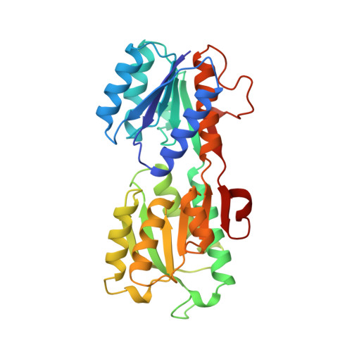
Structure of the periplasmic glucose/galactose receptor of Salmonella typhimurium.

Mowbray, S.L.Smith, R.D.Cole, L.B.

(1990) Receptor 1: 41-53

  • Primary Citation of Related Structures:  
    3GBP

  • PubMed Abstract: 

    The X-ray structure of the periplasmic glucose/galactose receptor (binding protein) of Salmonella typhimurium has been solved by the method of multiple isomorphous replacement and refined to a resolution of 2.4 A with an R-factor of 15.8%. The protein consists of two highly-similar structural domains with a bound sugar molecule trapped between. A combined sequence/structural approach is used to compare the structure of this protein to a similar arabinose receptor. The new sequence alignment obtained is used to analyze a number of features conserved for structural and functional reasons. This information allowed the prediction of similar features within the sequence of the analogous ribose receptor. Some of the consequences of the structures for chemotaxis and transport are discussed.


  • Organizational Affiliation

    Howard Hughes Medical Institute, Southwestern Medical Center, Dallas, TX 75235.


Macromolecules
Find similar proteins by:  (by identity cutoff)  |  3D Structure
Entity ID: 1
MoleculeChains Sequence LengthOrganismDetailsImage
GALACTOSE-BINDING PROTEIN307Salmonella enterica subsp. enterica serovar TyphimuriumMutation(s): 0 
UniProt
Find proteins for P23905 (Salmonella typhimurium (strain LT2 / SGSC1412 / ATCC 700720))
Explore P23905 
Go to UniProtKB:  P23905
Entity Groups  
Sequence Clusters30% Identity50% Identity70% Identity90% Identity95% Identity100% Identity
UniProt GroupP23905
Sequence Annotations
Expand
  • Reference Sequence
Small Molecules
Experimental Data & Validation

Experimental Data

  • Method: X-RAY DIFFRACTION
  • Resolution: 2.40 Å
  • R-Value Work: 0.158 
  • Space Group: C 1 2 1
Unit Cell:
Length ( Å )Angle ( ˚ )
a = 119.59α = 90
b = 37.28β = 123.37
c = 80.23γ = 90
Software Package:
Software NamePurpose
X-PLORrefinement

Structure Validation

View Full Validation Report


Ligand Structure Quality Assessment 


Entry History 

Deposition Data

  • Released Date: 1991-07-15 
  • Deposition Author(s): Mowbray, S.L.
  • This entry supersedes: 1GBP

Revision History  (Full details and data files)

  • Version 1.0: 1991-07-15
    Type: Initial release
  • Version 1.1: 2008-03-03
    Changes: Version format compliance
  • Version 1.2: 2011-07-13
    Changes: Version format compliance
  • Version 1.3: 2017-11-29
    Changes: Derived calculations, Other
  • Version 2.0: 2020-07-29
    Type: Remediation
    Reason: Carbohydrate remediation
    Changes: Advisory, Atomic model, Data collection, Database references, Derived calculations, Structure summary
  • Version 2.1: 2024-02-21
    Changes: Data collection, Database references, Structure summary