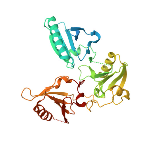
3FG6

Structure of the C-terminus of Adseverin


Experimental Data Snapshot

  • Method: X-RAY DIFFRACTION
  • Resolution: 3.00 Å
  • R-Value Free: 0.295 
  • R-Value Work: 0.244 
  • R-Value Observed: 0.247 

wwPDB Validation   3D Report Full Report


This is version 1.3 of the entry. See complete history


Literature

The crystal structure of the C-terminus of adseverin reveals the actin-binding interface.

Chumnarnsilpa, S.Lee, W.L.Nag, S.Kannan, B.Larsson, M.Burtnick, L.D.Robinson, R.C.

(2009) Proc Natl Acad Sci U S A 106: 13719-13724

  • DOI: https://doi.org/10.1073/pnas.0812383106
  • Primary Citation of Related Structures:  
    3FG6

  • PubMed Abstract: 

    Adseverin is a member of the calcium-regulated gelsolin superfamily of actin severing and capping proteins. Adseverin comprises 6 homologous domains (A1-A6), which share 60% identity with the 6 domains from gelsolin (G1-G6). Adseverin is truncated in comparison to gelsolin, lacking the C-terminal extension that masks the F-actin binding site in calcium-free gelsolin. Biochemical assays have indicated differences in the interaction of the C-terminal halves of adseverin and gelsolin with actin. Gelsolin contacts actin through a major site on G4 and a minor site on G6, whereas adseverin uses a site on A5. Here, we present the X-ray structure of the activated C-terminal half of adseverin (A4-A6). This structure is highly similar to that of the activated form of the C-terminal half of gelsolin (G4-G6), both in arrangement of domains and in the 3 bound calcium ions. Comparative analysis of the actin-binding surfaces observed in the G4-G6/actin structure suggests that adseverin in this conformation will also be able to interact with actin through A4 and A6, whereas the A5 surface is obscured. A single residue mutation in A4-A6 located at the predicted A4/actin interface completely abrogates actin sequestration. A model of calcium-free adseverin, constructed from the structure of gelsolin, predicts that in the absence of a gelsolin-like C-terminal extension the interaction between A2 and A6 provides the steric inhibition to prevent interaction with F-actin. We propose that calcium binding to the N terminus of adseverin dominates the activation process to expose the F-actin binding site on A2.


  • Organizational Affiliation

    Institute of Molecular and Cell Biology, Agency for Science, Technology, and Research (ASTAR), 61 Biopolis Drive, Proteos, Singapore 138673.


Macromolecules
Find similar proteins by:  (by identity cutoff)  |  3D Structure
Entity ID: 1
MoleculeChains Sequence LengthOrganismDetailsImage
Adseverin371Homo sapiensMutation(s): 0 
Gene Names: SCINKIAA1905
UniProt & NIH Common Fund Data Resources
Find proteins for Q9Y6U3 (Homo sapiens)
Explore Q9Y6U3 
Go to UniProtKB:  Q9Y6U3
PHAROS:  Q9Y6U3
GTEx:  ENSG00000006747 
Entity Groups  
Sequence Clusters30% Identity50% Identity70% Identity90% Identity95% Identity100% Identity
UniProt GroupQ9Y6U3
Sequence Annotations
Expand
  • Reference Sequence
Small Molecules
Ligands 1 Unique
IDChains Name / Formula / InChI Key2D Diagram3D Interactions
CA
Query on CA

Download Ideal Coordinates CCD File 
AA [auth D]
BA [auth D]
CA [auth B]
DA [auth B]
EA [auth B]
AA [auth D],
BA [auth D],
CA [auth B],
DA [auth B],
EA [auth B],
I [auth A],
J [auth A],
K [auth A],
L [auth C],
M [auth C],
N [auth C],
O [auth G],
P [auth G],
Q [auth G],
R [auth E],
S [auth E],
T [auth E],
U [auth F],
V [auth F],
W [auth H],
X [auth H],
Y [auth H],
Z [auth D]
CALCIUM ION
Ca
BHPQYMZQTOCNFJ-UHFFFAOYSA-N
Experimental Data & Validation

Experimental Data

  • Method: X-RAY DIFFRACTION
  • Resolution: 3.00 Å
  • R-Value Free: 0.295 
  • R-Value Work: 0.244 
  • R-Value Observed: 0.247 
  • Space Group: P 1
Unit Cell:
Length ( Å )Angle ( ˚ )
a = 67.644α = 88.79
b = 90.384β = 88.26
c = 98.839γ = 76.26
Software Package:
Software NamePurpose
DENZOdata reduction
SCALEPACKdata scaling
PHASERphasing
REFMACrefinement
PDB_EXTRACTdata extraction

Structure Validation

View Full Validation Report


Entry History 

Deposition Data

Revision History  (Full details and data files)

  • Version 1.0: 2009-08-11
    Type: Initial release
  • Version 1.1: 2011-07-13
    Changes: Advisory, Refinement description, Version format compliance
  • Version 1.2: 2017-11-01
    Changes: Refinement description
  • Version 1.3: 2023-11-01
    Changes: Data collection, Database references, Derived calculations, Refinement description