3FG6 | pdb_00003fg6

Structure of the C-terminus of Adseverin


Experimental Data Snapshot

  • Method: X-RAY DIFFRACTION
  • Resolution: 3.00 Å
  • R-Value Free: 
    0.295 (Depositor), 0.274 (DCC) 
  • R-Value Work: 
    0.244 (Depositor), 0.233 (DCC) 
  • R-Value Observed: 
    0.247 (Depositor) 

Starting Model: experimental
View more details

wwPDB Validation 3D Report Full Report

Validation slider image for 3FG6

This is version 1.3 of the entry. See complete history

Literature

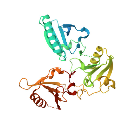
The crystal structure of the C-terminus of adseverin reveals the actin-binding interface.

Chumnarnsilpa, S.Lee, W.L.Nag, S.Kannan, B.Larsson, M.Burtnick, L.D.Robinson, R.C.

(2009) Proc Natl Acad Sci U S A 106: 13719-13724

  • DOI: https://doi.org/10.1073/pnas.0812383106
  • Primary Citation Related Structures: 
    3FG6

  • PubMed Abstract: 

    Adseverin is a member of the calcium-regulated gelsolin superfamily of actin severing and capping proteins. Adseverin comprises 6 homologous domains (A1-A6), which share 60% identity with the 6 domains from gelsolin (G1-G6). Adseverin is truncated in comparison to gelsolin, lacking the C-terminal extension that masks the F-actin binding site in calcium-free gelsolin. Biochemical assays have indicated differences in the interaction of the C-terminal halves of adseverin and gelsolin with actin. Gelsolin contacts actin through a major site on G4 and a minor site on G6, whereas adseverin uses a site on A5. Here, we present the X-ray structure of the activated C-terminal half of adseverin (A4-A6). This structure is highly similar to that of the activated form of the C-terminal half of gelsolin (G4-G6), both in arrangement of domains and in the 3 bound calcium ions. Comparative analysis of the actin-binding surfaces observed in the G4-G6/actin structure suggests that adseverin in this conformation will also be able to interact with actin through A4 and A6, whereas the A5 surface is obscured. A single residue mutation in A4-A6 located at the predicted A4/actin interface completely abrogates actin sequestration. A model of calcium-free adseverin, constructed from the structure of gelsolin, predicts that in the absence of a gelsolin-like C-terminal extension the interaction between A2 and A6 provides the steric inhibition to prevent interaction with F-actin. We propose that calcium binding to the N terminus of adseverin dominates the activation process to expose the F-actin binding site on A2.


  • Organizational Affiliation
    • Institute of Molecular and Cell Biology, Agency for Science, Technology, and Research (ASTAR), 61 Biopolis Drive, Proteos, Singapore 138673.

Macromolecule Content 

  • Total Structure Weight: 336.33 kDa 
  • Atom Count: 19,112 
  • Modeled Residue Count: 2,375 
  • Deposited Residue Count: 2,968 
  • Unique protein chains: 1

Macromolecules

Find similar proteins by:|  3D Structure
Entity ID: 1
MoleculeChains  Sequence LengthOrganismDetailsImage
Adseverin371Homo sapiensMutation(s): 0 
Gene Names: SCINKIAA1905
UniProt & NIH Common Fund Data Resources
Find proteins for Q9Y6U3 (Homo sapiens)
Explore Q9Y6U3 
Go to UniProtKB:  Q9Y6U3
PHAROS:  Q9Y6U3
GTEx:  ENSG00000006747 
Entity Groups
Sequence Clusters30% Identity50% Identity70% Identity90% Identity95% Identity100% Identity
UniProt GroupQ9Y6U3
Sequence Annotations
Expand
Reference Sequence

Small Molecules

Ligands 1 Unique
IDChains Name / Formula / InChI Key2D Diagram3D Interactions
CA

Query on CA


Download:Ideal Coordinates CCD File
AA [auth D]
BA [auth D]
CA [auth B]
DA [auth B]
EA [auth B]
AA [auth D],
BA [auth D],
CA [auth B],
DA [auth B],
EA [auth B],
I [auth A],
J [auth A],
K [auth A],
L [auth C],
M [auth C],
N [auth C],
O [auth G],
P [auth G],
Q [auth G],
R [auth E],
S [auth E],
T [auth E],
U [auth F],
V [auth F],
W [auth H],
X [auth H],
Y [auth H],
Z [auth D]
CALCIUM ION
Ca
BHPQYMZQTOCNFJ-UHFFFAOYSA-N

Experimental Data & Validation

Experimental Data

  • Method: X-RAY DIFFRACTION
  • Resolution: 3.00 Å
  • R-Value Free:  0.295 (Depositor), 0.274 (DCC) 
  • R-Value Work:  0.244 (Depositor), 0.233 (DCC) 
  • R-Value Observed: 0.247 (Depositor) 
Space Group: P 1
Unit Cell:
Length ( Å )Angle ( ˚ )
a = 67.644α = 88.79
b = 90.384β = 88.26
c = 98.839γ = 76.26
Software Package:
Software NamePurpose
DENZOdata reduction
SCALEPACKdata scaling
PHASERphasing
REFMACrefinement
PDB_EXTRACTdata extraction

Structure Validation

View Full Validation Report


Entry History 

Deposition Data

Revision History  (Full details and data files)

  • Version 1.0: 2009-08-11
    Type: Initial release
  • Version 1.1: 2011-07-13
    Changes: Advisory, Refinement description, Version format compliance
  • Version 1.2: 2017-11-01
    Changes: Refinement description
  • Version 1.3: 2023-11-01
    Changes: Data collection, Database references, Derived calculations, Refinement description