3DPG

SgrAI with noncognate DNA bound


Experimental Data Snapshot

  • Method: X-RAY DIFFRACTION
  • Resolution: 1.91 Å
  • R-Value Free: 0.225 
  • R-Value Work: 0.185 
  • R-Value Observed: 0.187 

wwPDB Validation   3D Report Full Report


This is version 1.3 of the entry. See complete history


Literature

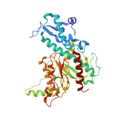
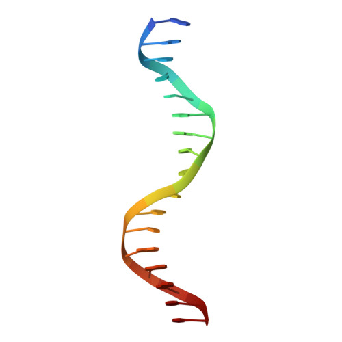
The structure of SgrAI bound to DNA; recognition of an 8 base pair target.

Dunten, P.W.Little, E.J.Gregory, M.T.Manohar, V.M.Dalton, M.Hough, D.Bitinaite, J.Horton, N.C.

(2008) Nucleic Acids Res 36: 5405-5416

  • DOI: https://doi.org/10.1093/nar/gkn510
  • Primary Citation of Related Structures:  
    3DPG, 3DVO, 3DW9

  • PubMed Abstract: 

    The three-dimensional X-ray crystal structure of the 'rare cutting' type II restriction endonuclease SgrAI bound to cognate DNA is presented. SgrAI forms a dimer bound to one duplex of DNA. Two Ca(2+) bind in the enzyme active site, with one ion at the interface between the protein and DNA, and the second bound distal from the DNA. These sites are differentially occupied by Mn(2+), with strong binding at the protein-DNA interface, but only partial occupancy of the distal site. The DNA remains uncleaved in the structures from crystals grown in the presence of either divalent cation. The structure of the dimer of SgrAI is similar to those of Cfr10I, Bse634I and NgoMIV, however no tetrameric structure of SgrAI is observed. DNA contacts to the central CCGG base pairs of the SgrAI canonical target sequence (CR|CCGGYG, | marks the site of cleavage) are found to be very similar to those in the NgoMIV/DNA structure (target sequence G|CCGGC). Specificity at the degenerate YR base pairs of the SgrAI sequence may occur via indirect readout using DNA distortion. Recognition of the outer GC base pairs occurs through a single contact to the G from an arginine side chain located in a region unique to SgrAI.


  • Organizational Affiliation

    Stanford Synchrotron Radiation Laboratory, Stanford University, Menlo Park, CA 94025, USA.


Macromolecules

Find similar proteins by:  (by identity cutoff)  |  3D Structure
Entity ID: 2
MoleculeChains Sequence LengthOrganismDetailsImage
SgraIR restriction enzymeC [auth A],
D [auth B]
338Streptomyces griseusMutation(s): 0 
Gene Names: sgraIR
EC: 3.1.21.4
UniProt
Find proteins for Q9F6L0 (Streptomyces griseus)
Explore Q9F6L0 
Go to UniProtKB:  Q9F6L0
Entity Groups  
Sequence Clusters30% Identity50% Identity70% Identity90% Identity95% Identity100% Identity
UniProt GroupQ9F6L0
Sequence Annotations
Expand
  • Reference Sequence

Find similar nucleic acids by:  Sequence   |   3D Structure  

Entity ID: 1
MoleculeChains LengthOrganismImage
DNA (5'-D(*DAP*DAP*DGP*DTP*DCP*DGP*DAP*DCP*DCP*DGP*DGP*DTP*DGP*DGP*DAP*DCP*DT)-3')A [auth C],
B [auth D]
17N/A
Sequence Annotations
Expand
  • Reference Sequence
Experimental Data & Validation

Experimental Data

  • Method: X-RAY DIFFRACTION
  • Resolution: 1.91 Å
  • R-Value Free: 0.225 
  • R-Value Work: 0.185 
  • R-Value Observed: 0.187 
  • Space Group: P 1 21 1
Unit Cell:
Length ( Å )Angle ( ˚ )
a = 69.726α = 90
b = 78.621β = 109.76
c = 86.834γ = 90
Software Package:
Software NamePurpose
PHENIXrefinement
MOSFLMdata reduction
SCALAdata scaling
DMphasing

Structure Validation

View Full Validation Report


Entry History 

Deposition Data

Revision History  (Full details and data files)

  • Version 1.0: 2008-08-19
    Type: Initial release
  • Version 1.1: 2011-07-13
    Changes: Version format compliance
  • Version 1.2: 2020-02-26
    Changes: Data collection, Database references
  • Version 1.3: 2024-02-21
    Changes: Data collection, Database references, Derived calculations