3D7W

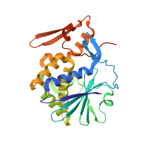
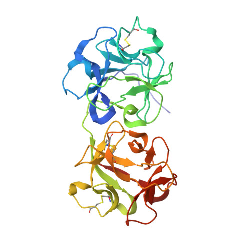
Mistletoe Lectin I in Complex with Zeatin


Experimental Data Snapshot

  • Method: X-RAY DIFFRACTION
  • Resolution: 2.49 Å
  • R-Value Free: 0.224 
  • R-Value Work: 0.182 
  • R-Value Observed: 0.184 

wwPDB Validation   3D Report Full Report


Ligand Structure Quality Assessment 


This is version 2.1 of the entry. See complete history


Literature

Structure of mistletoe lectin I from Viscum album in complex with the phytohormone zeatin

Meyer, A.Rypniewski, W.Szymanski, M.Voelter, W.Barciszewski, J.Betzel, C.

(2008) Biochim Biophys Acta 1784: 1590-1595

  • DOI: https://doi.org/10.1016/j.bbapap.2008.07.010
  • Primary Citation of Related Structures:  
    3D7W

  • PubMed Abstract: 

    The crystal structure of mistletoe lectin I (ML-I) isolated from the European mistletoe Viscum album in complex with the most active phytohormone zeatin has been analyzed and refined to 2.54 A resolution. X-ray suitable crystals of ML-I were obtained by the counter-diffusion method using the Gel-Tube R crystallization kit (GT-R) onboard the Russian Service Module on the international space station ISS. High quality hexagonal bipyramidal crystals were grown during 3 months under microgravity conditions. Selected crystals were soaked in a saturated solution of zeatin and subsequently diffraction data were collected applying synchrotron radiation. A distinct F(o)-F(c) electron density has been found inside a binding pocket located in subunit B of ML-I and has been interpreted as a single zeatin molecule. The structure was refined to investigate the zeatin-ML-I interactions in detail. The results demonstrate the ability of mistletoe to protect itself from the host transpiration regulation by absorbing the most active host plant hormones as part of a defense mechanism.


  • Organizational Affiliation

    Institute of Biochemistry and Molecular Biology, University of Hamburg, c/o DESY, Notkestr. 85, Building 22a, 22603 Hamburg, Germany.


Macromolecules
Find similar proteins by:  (by identity cutoff)  |  3D Structure
Entity ID: 1
MoleculeChains Sequence LengthOrganismDetailsImage
Beta-galactoside-specific lectin 1254Viscum albumMutation(s): 0 
EC: 3.2.2.22
UniProt
Find proteins for P81446 (Viscum album)
Explore P81446 
Go to UniProtKB:  P81446
Entity Groups  
Sequence Clusters30% Identity50% Identity70% Identity90% Identity95% Identity100% Identity
UniProt GroupP81446
Sequence Annotations
Expand
  • Reference Sequence
Find similar proteins by:  (by identity cutoff)  |  3D Structure
Entity ID: 2
MoleculeChains Sequence LengthOrganismDetailsImage
Beta-galactoside-specific lectin 1265Viscum albumMutation(s): 0 
EC: 3.2.2.22
UniProt
Find proteins for P81446 (Viscum album)
Explore P81446 
Go to UniProtKB:  P81446
Entity Groups  
Sequence Clusters30% Identity50% Identity70% Identity90% Identity95% Identity100% Identity
UniProt GroupP81446
Sequence Annotations
Expand
  • Reference Sequence
Oligosaccharides

Help

Entity ID: 3
MoleculeChains Length2D Diagram Glycosylation3D Interactions
2-acetamido-2-deoxy-beta-D-glucopyranose-(1-4)-2-acetamido-2-deoxy-beta-D-glucopyranose
C, D
2N-Glycosylation
Glycosylation Resources
GlyTouCan:  G42666HT
GlyCosmos:  G42666HT
GlyGen:  G42666HT
Small Molecules
Ligands 5 Unique
IDChains Name / Formula / InChI Key2D Diagram3D Interactions
NAG
Query on NAG

Download Ideal Coordinates CCD File 
E [auth A],
J [auth B]
2-acetamido-2-deoxy-beta-D-glucopyranose
C8 H15 N O6
OVRNDRQMDRJTHS-FMDGEEDCSA-N
ZEZ
Query on ZEZ

Download Ideal Coordinates CCD File 
L [auth B](2Z)-2-methyl-4-(9H-purin-6-ylamino)but-2-en-1-ol
C10 H13 N5 O
UZKQTCBAMSWPJD-UQCOIBPSSA-N
ZEA
Query on ZEA

Download Ideal Coordinates CCD File 
K [auth B](2E)-2-methyl-4-(9H-purin-6-ylamino)but-2-en-1-ol
C10 H13 N5 O
UZKQTCBAMSWPJD-FARCUNLSSA-N
SO4
Query on SO4

Download Ideal Coordinates CCD File 
F [auth A],
G [auth A]
SULFATE ION
O4 S
QAOWNCQODCNURD-UHFFFAOYSA-L
GOL
Query on GOL

Download Ideal Coordinates CCD File 
H [auth A],
I [auth A],
M [auth B],
N [auth B]
GLYCEROL
C3 H8 O3
PEDCQBHIVMGVHV-UHFFFAOYSA-N
Experimental Data & Validation

Experimental Data

  • Method: X-RAY DIFFRACTION
  • Resolution: 2.49 Å
  • R-Value Free: 0.224 
  • R-Value Work: 0.182 
  • R-Value Observed: 0.184 
  • Space Group: P 65 2 2
Unit Cell:
Length ( Å )Angle ( ˚ )
a = 106.644α = 90
b = 106.644β = 90
c = 311.667γ = 120
Software Package:
Software NamePurpose
REFMACrefinement

Structure Validation

View Full Validation Report


Ligand Structure Quality Assessment 


Entry History 

Deposition Data

Revision History  (Full details and data files)

  • Version 1.0: 2008-06-17
    Type: Initial release
  • Version 1.1: 2011-07-13
    Changes: Non-polymer description, Version format compliance
  • Version 1.2: 2014-02-05
    Changes: Derived calculations
  • Version 2.0: 2020-07-29
    Type: Remediation
    Reason: Carbohydrate remediation
    Changes: Advisory, Atomic model, Data collection, Derived calculations, Structure summary
  • Version 2.1: 2023-11-01
    Changes: Data collection, Database references, Refinement description, Structure summary