3LFM

Crystal structure of the fat mass and obesity associated (FTO) protein reveals basis for its substrate specificity


Experimental Data Snapshot

  • Method: X-RAY DIFFRACTION
  • Resolution: 2.50 Å
  • R-Value Free: 0.285 
  • R-Value Work: 0.239 

wwPDB Validation   3D Report Full Report


Ligand Structure Quality Assessment 


This is version 1.2 of the entry. See complete history


Literature

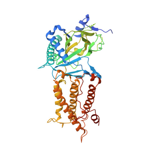
Crystal structure of the FTO protein reveals basis for its substrate specificity

Han, Z.Niu, T.Chang, J.Lei, X.Zhao, M.Wang, Q.Cheng, W.Wang, J.Feng, Y.Chai, J.

(2010) Nature 464: 1205-1209

  • DOI: https://doi.org/10.1038/nature08921
  • Primary Citation of Related Structures:  
    3LFM

  • PubMed Abstract: 

    Recent studies have unequivocally associated the fat mass and obesity-associated (FTO) gene with the risk of obesity. In vitro FTO protein is an AlkB-like DNA/RNA demethylase with a strong preference for 3-methylthymidine (3-meT) in single-stranded DNA or 3-methyluracil (3-meU) in single-stranded RNA. Here we report the crystal structure of FTO in complex with the mononucleotide 3-meT. FTO comprises an amino-terminal AlkB-like domain and a carboxy-terminal domain with a novel fold. Biochemical assays show that these two domains interact with each other, which is required for FTO catalytic activity. In contrast with the structures of other AlkB members, FTO possesses an extra loop covering one side of the conserved jelly-roll motif. Structural comparison shows that this loop selectively competes with the unmethylated strand of the DNA duplex for binding to FTO, suggesting that it has an important role in FTO selection against double-stranded nucleic acids. The ability of FTO to distinguish 3-meT or 3-meU from other nucleotides is conferred by its hydrogen-bonding interaction with the two carbonyl oxygen atoms in 3-meT or 3-meU. Taken together, these results provide a structural basis for understanding FTO substrate-specificity, and serve as a foundation for the rational design of FTO inhibitors.


  • Organizational Affiliation

    National Institute of Biological Sciences, No. 7 Science Park Road, Beijing 102206, China.


Macromolecules
Find similar proteins by:  (by identity cutoff)  |  3D Structure
Entity ID: 1
MoleculeChains Sequence LengthOrganismDetailsImage
Protein fto495Homo sapiensMutation(s): 0 
Gene Names: FTO
EC: 1.14.11
UniProt & NIH Common Fund Data Resources
Find proteins for Q9C0B1 (Homo sapiens)
Explore Q9C0B1 
Go to UniProtKB:  Q9C0B1
PHAROS:  Q9C0B1
GTEx:  ENSG00000140718 
Entity Groups  
Sequence Clusters30% Identity50% Identity70% Identity90% Identity95% Identity100% Identity
UniProt GroupQ9C0B1
Sequence Annotations
Expand
  • Reference Sequence
Small Molecules
Binding Affinity Annotations 
IDSourceBinding Affinity
OGA BindingDB:  3LFM IC50: 4.40e+4 (nM) from 1 assay(s)
Experimental Data & Validation

Experimental Data

  • Method: X-RAY DIFFRACTION
  • Resolution: 2.50 Å
  • R-Value Free: 0.285 
  • R-Value Work: 0.239 
  • Space Group: H 3
Unit Cell:
Length ( Å )Angle ( ˚ )
a = 142.86α = 90
b = 142.86β = 90
c = 83.8γ = 120
Software Package:
Software NamePurpose
HKL-2000data collection
SOLVEphasing
CNSrefinement
DENZOdata reduction
SCALEPACKdata scaling

Structure Validation

View Full Validation Report


Ligand Structure Quality Assessment 


Entry History 

Deposition Data

Revision History  (Full details and data files)

  • Version 1.0: 2010-04-07
    Type: Initial release
  • Version 1.1: 2011-07-13
    Changes: Version format compliance
  • Version 1.2: 2024-03-20
    Changes: Data collection, Database references, Derived calculations