3G9R

Structure of the HIV-1 gp41 Membrane-Proximal Ectodomain Region in a Putative Prefusion Conformation


Experimental Data Snapshot

  • Method: X-RAY DIFFRACTION
  • Resolution: 2.00 Å
  • R-Value Free: 0.269 
  • R-Value Work: 0.213 
  • R-Value Observed: 0.216 

wwPDB Validation   3D Report Full Report


This is version 1.4 of the entry. See complete history


Literature

Structure of the HIV-1 gp41 Membrane-Proximal Ectodomain Region in a Putative Prefusion Conformation.

Liu, J.Deng, Y.Dey, A.Moore, J.Lu, M.

(2009) Biochemistry 48: 2915-2923

  • DOI: https://doi.org/10.1021/bi802303b
  • Primary Citation of Related Structures:  
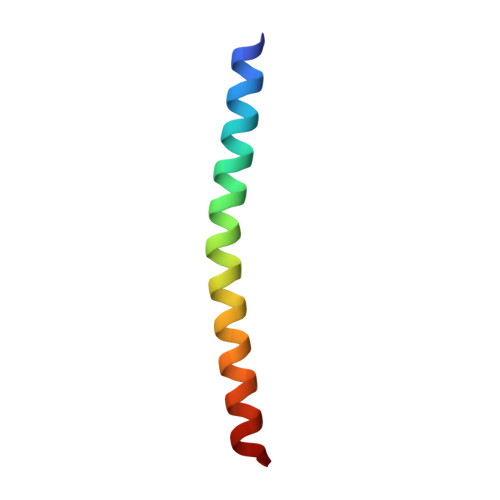
    3G9R

  • PubMed Abstract: 

    The conserved membrane-proximal external region (MPER) of the HIV-1 gp41 envelope protein is the established target for very rare but broadly neutralizing monoclonal antibodies (NAbs) elicited during natural human infection. Nevertheless, attempts to generate an HIV-1 neutralizing antibody response with immunogens bearing MPER epitopes have met with limited success. Here we show that the MPER peptide (residues 662-683) forms a labile alpha-helical trimer in aqueous solution and report the crystal structure of this autonomous folding subdomain stabilized by addition of a C-terminal isoleucine zipper motif. The structure reveals a parallel triple-stranded coiled coil in which the neutralization epitope residues are buried within the interface between the associating MPER helices. Accordingly, both the 2F5 and 4E10 NAbs recognize the isolated MPER peptide but fail to bind the trimeric MPER subdomain. We propose that the trimeric MPER structure represents the prefusion conformation of gp41, preceding the putative prehairpin intermediate and the postfusion trimer-of-hairpins structure. As such, the MPER trimer should inform the design of new HIV-1 immunogens to elicit broadly neutralizing antibodies.


  • Organizational Affiliation

    Department of Biochemistry, Weill Medical College of Cornell University, New York, New York 10021, USA.


Macromolecules
Find similar proteins by:  (by identity cutoff)  |  3D Structure
Entity ID: 1
MoleculeChains Sequence LengthOrganismDetailsImage
Fusion complex of HIV-1 Envelope glycoprotein and Saccharomyces cerevisiae General control protein GCN4
A, B, C, D, E
A, B, C, D, E, F
42Human immunodeficiency virus 1Saccharomyces cerevisiae
This entity is chimeric
Mutation(s): 8 
UniProt
Find proteins for P03069 (Saccharomyces cerevisiae (strain ATCC 204508 / S288c))
Explore P03069 
Go to UniProtKB:  P03069
Find proteins for P03377 (Human immunodeficiency virus type 1 group M subtype B (isolate BRU/LAI))
Explore P03377 
Go to UniProtKB:  P03377
Entity Groups  
Sequence Clusters30% Identity50% Identity70% Identity90% Identity95% Identity100% Identity
UniProt GroupsP03069P03377
Sequence Annotations
Expand
  • Reference Sequence
Small Molecules
Ligands 2 Unique
IDChains Name / Formula / InChI Key2D Diagram3D Interactions
MPD
Query on MPD

Download Ideal Coordinates CCD File 
I [auth A]
J [auth A]
K [auth A]
L [auth A]
N [auth B]
I [auth A],
J [auth A],
K [auth A],
L [auth A],
N [auth B],
P [auth C],
R [auth E],
S [auth E]
(4S)-2-METHYL-2,4-PENTANEDIOL
C6 H14 O2
SVTBMSDMJJWYQN-YFKPBYRVSA-N
PO4
Query on PO4

Download Ideal Coordinates CCD File 
G [auth A],
H [auth A],
M [auth B],
O [auth C],
Q [auth D]
PHOSPHATE ION
O4 P
NBIIXXVUZAFLBC-UHFFFAOYSA-K
Experimental Data & Validation

Experimental Data

  • Method: X-RAY DIFFRACTION
  • Resolution: 2.00 Å
  • R-Value Free: 0.269 
  • R-Value Work: 0.213 
  • R-Value Observed: 0.216 
  • Space Group: P 1 21 1
Unit Cell:
Length ( Å )Angle ( ˚ )
a = 42.527α = 90
b = 48.157β = 95.38
c = 81.782γ = 90
Software Package:
Software NamePurpose
ADSCdata collection
AMoREphasing
REFMACrefinement
DENZOdata reduction
SCALEPACKdata scaling

Structure Validation

View Full Validation Report


Entry History 

Deposition Data

  • Released Date: 2009-05-12 
  • Deposition Author(s): Liu, J., Lu, M.

Revision History  (Full details and data files)

  • Version 1.0: 2009-05-12
    Type: Initial release
  • Version 1.1: 2011-07-13
    Changes: Version format compliance
  • Version 1.2: 2017-08-02
    Changes: Refinement description, Source and taxonomy
  • Version 1.3: 2021-10-20
    Changes: Database references, Derived calculations
  • Version 1.4: 2023-09-06
    Changes: Data collection, Refinement description