2Z4T

Crystal Structure of Vibrionaceae Photobacterium sp. JT-ISH-224 2,6-sialyltransferase in a Ternary Complex with Donor Product CMP and Accepter Substrate Lactose


Experimental Data Snapshot

  • Method: X-RAY DIFFRACTION
  • Resolution: 2.50 Å
  • R-Value Free: 0.243 
  • R-Value Work: 0.192 
  • R-Value Observed: 0.195 

wwPDB Validation   3D Report Full Report


Ligand Structure Quality Assessment 


This is version 2.1 of the entry. See complete history


Literature

Crystal structure of Vibrionaceae Photobacterium sp. JT-ISH-224 alpha2,6-sialyltransferase in a ternary complex with donor product CMP and acceptor substrate lactose: catalytic mechanism and substrate recognition

Kakuta, Y.Okino, N.Kajiwara, H.Ichikawa, M.Takakura, Y.Ito, M.Yamamoto, T.

(2008) Glycobiology 18: 66-73

  • DOI: https://doi.org/10.1093/glycob/cwm119
  • Primary Citation of Related Structures:  
    2Z4T

  • PubMed Abstract: 

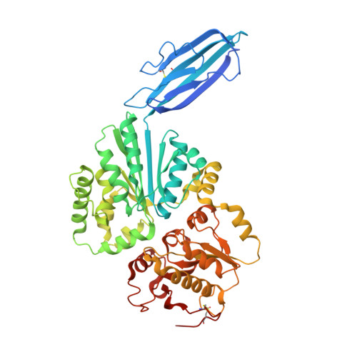
    Sialyltransferases are a family of glycosyltransferases that catalyze the transfer of N-acetylneuraminic acid residues from cytidine monophosphate N-acetylneuraminic acid (CMP-NeuAc) as a donor substrate to the carbohydrate groups of glycoproteins and glycolipids as acceptor substrates. We determined the crystal structure of Delta16psp26ST, the N-terminal truncated form of alpha2,6-sialyltransferase from Vibrionaceae Photobacterium sp. JT-ISH-224, complexed with a donor product CMP and an acceptor substrate lactose. Delta16psp26ST has three structural domains. Domain 1 belongs to the immunoglobulin-like beta-sandwich fold, and domains 2 and 3 form the glycosyltransferase-B structure. The CMP and lactose were bound in the deep cleft between domains 2 and 3. In the structure, only Asp232 was within hydrogen-binding distance of the acceptor O6 carbon of the galactose residue in lactose, and His405 was within hydrogen-binding distance of the phosphate oxygen of CMP. Mutation of these residues greatly decreased the activity of the enzyme. These structural and mutational results indicated that Asp232 might act as a catalytic base for deprotonation of the acceptor substrate, and His405 might act as a catalytic acid for protonation of the donor substrate. These findings are consistent with an in-line-displacement reaction mechanism in which Delta16psp26ST catalyzes the inverting transfer reaction. Unlike the case with multifunctional sialyltransferase (Delta24PmST1) complexed with CMP and lactose, the crystal structure of which was recently reported, the alpha2,6 reaction specificity of Delta16psp26ST is likely to be determined by His123.


  • Organizational Affiliation

    Department of Bioscience and Biotechnology, Graduate School of Bioresource and Bioenvironmental Sciences, Kyushu University, 6-10-1 Hakozaki, Higashi-ku, Fukuoka 812-8581, Japan. kakuta@agr.kyushu-u.ac.jp


Macromolecules
Find similar proteins by:  (by identity cutoff)  |  3D Structure
Entity ID: 1
MoleculeChains Sequence LengthOrganismDetailsImage
Beta-galactoside alpha-2,6-sialyltransferase514Photobacterium sp. JT-ISH-224Mutation(s): 0 
Gene Names: pst6-224
UniProt
Find proteins for A8QYL1 (Photobacterium sp. JT-ISH-224)
Explore A8QYL1 
Go to UniProtKB:  A8QYL1
Entity Groups  
Sequence Clusters30% Identity50% Identity70% Identity90% Identity95% Identity100% Identity
UniProt GroupA8QYL1
Sequence Annotations
Expand
  • Reference Sequence
Oligosaccharides

Help

Entity ID: 2
MoleculeChains Length2D Diagram Glycosylation3D Interactions
beta-D-galactopyranose-(1-4)-alpha-D-glucopyranose
B
2N/A
Glycosylation Resources
GlyTouCan:  G88362QR
GlyCosmos:  G88362QR
GlyGen:  G88362QR
Small Molecules
Ligands 4 Unique
IDChains Name / Formula / InChI Key2D Diagram3D Interactions
C5P
Query on C5P

Download Ideal Coordinates CCD File 
C [auth A]CYTIDINE-5'-MONOPHOSPHATE
C9 H14 N3 O8 P
IERHLVCPSMICTF-XVFCMESISA-N
PO4
Query on PO4

Download Ideal Coordinates CCD File 
I [auth A],
J [auth A]
PHOSPHATE ION
O4 P
NBIIXXVUZAFLBC-UHFFFAOYSA-K
GOL
Query on GOL

Download Ideal Coordinates CCD File 
D [auth A],
E [auth A],
F [auth A],
G [auth A],
H [auth A]
GLYCEROL
C3 H8 O3
PEDCQBHIVMGVHV-UHFFFAOYSA-N
MG
Query on MG

Download Ideal Coordinates CCD File 
K [auth A],
L [auth A],
M [auth A]
MAGNESIUM ION
Mg
JLVVSXFLKOJNIY-UHFFFAOYSA-N
Biologically Interesting Molecules (External Reference) 1 Unique
Experimental Data & Validation

Experimental Data

  • Method: X-RAY DIFFRACTION
  • Resolution: 2.50 Å
  • R-Value Free: 0.243 
  • R-Value Work: 0.192 
  • R-Value Observed: 0.195 
  • Space Group: P 31 2 1
Unit Cell:
Length ( Å )Angle ( ˚ )
a = 90.292α = 90
b = 90.292β = 90
c = 204.33γ = 120
Software Package:
Software NamePurpose
REFMACrefinement
HKL-2000data reduction
HKL-2000data scaling
MOLREPphasing

Structure Validation

View Full Validation Report


Ligand Structure Quality Assessment 


Entry History 

Deposition Data

Revision History  (Full details and data files)

  • Version 1.0: 2008-04-08
    Type: Initial release
  • Version 1.1: 2011-07-13
    Changes: Advisory, Source and taxonomy, Version format compliance
  • Version 2.0: 2020-07-29
    Type: Remediation
    Reason: Carbohydrate remediation
    Changes: Advisory, Atomic model, Data collection, Derived calculations, Non-polymer description, Structure summary
  • Version 2.1: 2023-11-01
    Changes: Advisory, Data collection, Database references, Refinement description, Structure summary